
Peer review process
Not revised: This Reviewed Preprint includes the authors’ original preprint (without revision), an eLife assessment, public reviews, and a provisional response from the authors.
Read more about eLife’s peer review process.Editors
- Reviewing EditorVirginie Courtier-OrgogozoCNRS - Universite Paris Cite, Paris, France
- Senior EditorAlbert CardonaUniversity of Cambridge, Cambridge, United Kingdom
Reviewer #1 (Public Review):
Summary:
In this paper, Li and colleagues have found mircoRNAs that affect levels of metamorphosis-regulating genes that can also affect levels of sesquiterpenoids (juvenile hormone and related compounds) and ecdysteriods, which regulate the timing and stages of insects, respectively. They first compared the transcriptomes of Drosophila at the third larval instar and at the white pre-pupa stage. They found thousands of differences in gene transcript levels between males and females, and between the two different stages. Among those genes that were differentially regulated they saw that genes involved in insect hormone biosynthesis were disproportionately represented. Many of the differentially regulated genes were involved in the insect hormone biosynthesis pathway and ascorbate and alderete metabolism. MicroRNAs were also differentially expressed during metamorphosis and were separately identified. The authors then considered genes and whether the differentially expressed microRNAs might regulate transcripts known to be involved in sesquiterpenoid production. In silico analysis of microRNAs predicted a list of 17 microRNAs that can regulate transcripts of sesquiterpenoid biosynthesis genes. The authors then used an in vitro luciferase assay to validate the binding and downregulation of 10 of the microRNAs to genes involved with sesquiterpenoid production in S2 cells.
Li and colleagues then focus on two genes they found were bound by microRNAs that have established roles in metamorphosis. The microRNAs miR-34 and miR-277 bind transcripts of two protein-coding genes that regulate metamorphosis Kr-h1, which encodes a transcription factor that is a JH-inducible transcription factor, and Allatostatin C Receptor 1, (AstC-R1), a G-protein coupled receptor that regulates the corpora allatum, the gland that produces sesquiterpenoids. Using a LAMP assay, one of the microRNAs, miR-277 was shown to bind to both AstC-R1 and Kr-h1 in in vivo whole-animal extracts. There is no mention of binding between either protein-coding transcript and the miR-34 microRNA. Temporal expression of all four transcripts shows that their abundance is anti-correlated; stages of high miR-34 or miR-277 expression correlate with low AstC-R1 or Kr-h1 expression. Homozygous deletions of both mircroRNAs result in 23% lethality, five days after adult eclosion. The authors also generated specific mutants in miR-34 or miR-277 and find differences in the expression of AstC-R1 and Kr-h1 and sex-specific differences in both sesquiterpenoids and ecdysteroids in the knock-out lines. If there were phenotypes associated with the specific knock-outs, those were not mentioned. Next, the authors examined the transcriptomes of the miR-3277 and miR-34 mutants and found several other GO-terms enriched among the differentially expressed genes. However, the sesquiterpenoid pathway and ascorbate and alderete metabolism are not listed.
Strengths:
This is an interesting manuscript that could make an important contribution to our understanding of the roles of micro RNAs at metamorphosis, and potentially of how sex-specific differences arise during metamorphosis. Strengths of the paper include the functional validation of microRNA binding, in vitro and in vivo-, as well as the characterization of sesquiterpenoid and ecdysteroid titers. The authors have also used CRISPR to generate specific knock-outs of miR-34 and miR-277. The transcriptomes will be a resource for future work to mine for differences in gene expression during metamorphosis.
Weaknesses:
(1) Spatial Expression of miR-34 and miR-277. If miR-34 and miR-277 regulate AstC-R1 and Kr-h1, then they must be expressed in the same cells. Although the authors show that the microRNAs do bind to the transcripts of AstC-R1 and Kr-h1 in S2 cells, and miR-277 binds AstC-R1 and Kr-h1 in vivo whole-animal homogenates, we do not know if the microRNAs are ever in the cells where AstC-R1 or Kr-h1 are expressed. AstC-R1 is only expressed in a few cells in the brain, so it is not at all certain that it is co-expressed with either microRNA. The creation of enhancer lines or in situ hybridization in Drosophila is straightforward and would sort this out.
(2) Phenotypes. Although a double deletion was used and specific knock-outs of both miR-34 and miR-277 were generated, the analysis of the mutants is very superficial. For the homozygous deletion of both microRNAs miR-34 and miR-277, only a decrease in survivorship was observed a full six days after adult eclosion - after the end of metamorphosis. No phenotype for either miR-34KO or miR-277-KO was given. The authors cite the work of others who have found specific phenotypes after manipulation of sesquiterpenoids or ecdysteroids, like Riddiford and Ashburner, but do not use any of these many studies to help them characterize the phenotype. If the loss of miR-34 and miR-277 affects so many pathways (including MAPK signaling, TGF-beta signaling, FoxO signaling, and Wnt signaling), as well as global titers of metamorphic hormones, then there shouldn't there be something different in the development to discuss?
(3) I think the reliance on GO term enrichment is getting in the way of biology. For instance, I would not describe Kr-h1 as a sesquiterpenoid biosynthesis pathway gene. Yet the authors say they were motivated to examine microRNA regulation of Kr-h1 because they saw differences in levels of the sesquiterpenoid biosynthesis pathway between WL3 and WPP, a period which also saw differences in expression of some microRNAs. I understand that Kr-h1 expression is regulated by JH, a sesquiterpenoid, but it is not directly involved with JH production, so relying on GO term enrichment has made the decision to focus on Kr-h1 feel arbitrary.
(4) The transcriptomes of miR-34 and miR-277 should have revealed genes encoding members of the sesquiterpenoid biosynthesis pathway as well as AstC-R1 and Kr-h1, but neither was mentioned. The functional tests of miR-34 and miR-277 were performed because they were shown to affect the levels of expression of genes in the sesquiterpenoid biosynthesis pathway. Figure 2 shows a significant decrease in AstC-R1 and Kr-h1 transcripts after the loss of miR-34 and miR-277. However, the results do not mention either (Lines 250-264). Instead, there is a list of 10 different GO terms (like arginine and proline metabolism or fatty acid degradation) that were enriched in miR-34 and miR-277 transcriptomes. If any of those ten types have any relationship to Kr-h1, AstC-R1, or metamorphosis, that has not been explained.
(5) Not enough care was taken in describing the stages. The methods describe wandering larvae (WL3) and white pre-pupa (WPP) for the transcriptomes, but in the text, different terms are used, like "larva", "pupa" and "L3 larvae instars" "early pupae" "late L3". Also, it seems like the small RNA libraries for sequencing were taken from "L3 larvae", but the stage of the L3 larvae was not mentioned. Staging is important, especially during metamorphosis, since differences in expression are expected to exist between different stages of L3, between early vs late wandering, and between WPP and early pupal stages.
Reviewer #2 (Public Review):
Summary:
This study proposes that the microRNA cluster miR-277/34 controls the generation of sexual dimorphism in Drosophila melanogaster during metamorphosis by acting on specific hormonal and developmental gene pathways.
Strengths:
Using a combination of mRNA and small RNA sequencing together with genome-wide in silico and in vitro analyses the authors identified a microRNA cluster that may be involved in metamorphosis and the generation of sexual dimorphism in Drosophila melanogaster.
Weaknesses:
Biological validation of the identified sexually dimorphic genes and a detailed understanding of how the microRNA cluster miR-277/34 might be involved in the regulation of sesquiterpenoids are needed.
Major suggestions:
(1) If AstC-R1 and Kr-h1 are targets of the miR-277/34 cluster and cause their downregulation, it is not clear why there would also be a decrease in the levels of these genes in the miR-277/34 mutants. This would suggest that the mechanism is not straightforward and that further epistatic experiments should be carried out in order to clarify this issue.
(2) The changes in the expression levels of AstC-R1 in pupae of miR-277-KO and mir-34-KO flies must be accompanied by photos of the respective larvae and pupae, as well as an analysis of the larvae-pupa transition on the mutants by gender.
(3) Biological validation of the identified sexually dimorphic genes in vivo will be necessary for the support of this work.
Reviewer #3 (Public Review):
Summary:
The authors show convincingly the complexity of gene up- and down-regulation at the outset of metamorphosis and identify substantial differences between the two sexes, even at this early time in development. The complexity of microRNA expression and the difference between the sexes are also nicely laid out. The functional significance of these differences, though, is harder to establish. The authors have focused on the roles of two families of developmental hormones, the ecdysteroids, and the juvenile hormones. The emergence of sex-specific differentiation of organs during metamorphosis is clearly downstream of the action of ecdysteroids and/or JH, but there is no evidence that the presence or lack of these hormones has any effect on the sexual identity of organ systems - i.e., that manipulations of JH or ecdysteroid result in either the masculinization or feminization of individuals or their organs. The precedence for the linkage of these hormones to sex determination is the 2002, Belgacem & Martin study, which describes the effects of JH on fly locomotion. These authors show that the number of stop/start bouts is sexually dimorphic, and removal of JH in males shifts their frequency into the female range while giving treated males exogenous JH moves it back. While this is referred to as a "feminization" of male behavior, this quantitative shift in frequency is not as compelling as would be a qualitative shift -- for example, the removal of JH causing males to show egg-laying behavior (a result that has never been seen). Also, these effects are in a fully mature system, rather than at the early metamorphic time examined in the present paper. In driving and coordination metamorphosis, JH and ecdysteroids are intimately involved in sexual differentiation, but I know of no compelling evidence that they play a role in sex determination.
While the summary of the effects or removal of specific microRNAs on the components of the biosynthetic pathway for the JHs and ecdysteroids (Figure 2E ,F) is quite compelling, I am concerned about the effects of the removal of mir-277 and mir-34 on the levels of both the JHs and 20E. My concern centers around the data from the control group (w[1118] animals in Figure 2D). These data are the first report of a marked sex difference in the titer of either JH or ecdysteroid at the start of metamorphosis in Drosophila. As expected, males show a 10-20 increase in levels of JH III, JHB3, and 20E between the L3 stage and the white puparium, but, surprisingly, the levels of these hormones in female L3 larvae are equal to or greater than that seen at pupariation! These data for females run counter to over 50 years of work on the effects of ecdysteroids in Drosophila!
As far as I can gather from the paper, the L3 data were obtained using wandering larvae. This stage lasts for about 12 hours and ends with pupariation. Larvae from this period need to be used with caution for hormone studies. Levels of both JH and ecdysteroid are low as larvae leave the food but rapidly rise to their peak levels at the white puparium stage 12 hours later. To deal with the rapidly changing hormonal landscape through this period, the researchers have used physiological markers to track this progression. Initially, it was the sage of "puffing" of the giant salivary gland chromosomes, but, for bulk collection of staged larvae, larvae are fed on food containing a blue dye, and progression is tracked by the loss of blue coloring from the gut. I could not find if the authors had any criteria for selecting larvae during the wandering period. Male and female larvae grow to different sizes. Might this difference in growth be biased when larvae were selected during their wandering phase?
The other hormone-related issue is the expression of Kr-h1 during larval stages and metamorphosis (Figure 1G). Kr-h1 is the main target of JH and Kr-h1 expression is often used as a proxy for the JH titer. The authors report that peak Kr-h1 expression occurs in the L3 (when the JH titer should be lowest!) and that it drops at wandering. This pattern is counter to that reported in the literature (e.g., FlyBase, ModEncode).
Author response:
[Editors’ note: An Expression of Concern has been published for this article. The authors’ responses submitted during revision have been appended for further context]
eLife Assessment
This study provides a useful inventory of genes that are up- or down-regulated during the early metamorphic development of male and female larvae and proposes that the microRNA cluster miR-277/34 is involved in the development of sexual differences during early metamorphosis of Drosophila melanogaster, although its precise role remains unclear. The strength of evidence, based on a combination of diverse methods including mRNA and small RNA sequencing, in silico analyses, in vitro assays, and loss-of-function experiments, is incomplete as it lacks a general model and an examination of the potential effects of the miR-277/34 mutations on phenotypes such as morphology or developmental time. This work will be of interest to developmental biologists interested in sexual dimorphism and in the interplay between hormones and microRNAs during development.
We thank the editor and reviewers for the positive and constructive comments and suggestions. Point-to-point responses are listed below.
Reviewer #1:
Summary:
In this paper, Li and colleagues have found mircoRNAs that affect levels of metamorphosis-regulating genes that can also affect levels of sesquiterpenoids (juvenile hormone and related compounds) and ecdysteriods, which regulate the timing and stages of insects, respectively. They first compared the transcriptomes of Drosophila at the third larval instar and at the white pre-pupa stage. They found thousands of differences in gene transcript levels between males and females, and between the two different stages. Among those genes that were differentially regulated they saw that genes involved in insect hormone biosynthesis were disproportionately represented. Many of the differentially regulated genes were involved in the insect hormone biosynthesis pathway and ascorbate and alderete metabolism. MicroRNAs were also differentially expressed during metamorphosis and were separately identified. The authors then considered genes and whether the differentially expressed microRNAs might regulate transcripts known to be involved in sesquiterpenoid production. In silico analysis of microRNAs predicted a list of 17 microRNAs that can regulate transcripts of sesquiterpenoid biosynthesis genes. The authors then used an in vitro luciferase assay to validate the binding and downregulation of 10 of the microRNAs to genes involved with sesquiterpenoid production in S2 cells.
Li and colleagues then focus on two genes they found were bound by microRNAs that have established roles in metamorphosis. The microRNAs miR-34 and miR-277 bind transcripts of two protein-coding genes that regulate metamorphosis Kr-h1, which encodes a transcription factor that is a JH-inducible transcription factor, and Allatostatin C Receptor 1, (AstC-R1), a G-protein coupled receptor that regulates the corpora allatum, the gland that produces sesquiterpenoids. Using a LAMP assay, one of the microRNAs, miR-277 was shown to bind to both AstC-R1 and Kr-h1 in in vivo whole-animal extracts. There is no mention of binding between either protein-coding transcript and the miR-34 microRNA. Temporal expression of all four transcripts shows that their abundance is anti-correlated; stages of high miR-34 or miR-277 expression correlate with low AstC-R1 or Kr-h1 expression. Homozygous deletions of both mircroRNAs result in 23% lethality, five days after adult eclosion. The authors also generated specific mutants in miR-34 or miR-277 and find differences in the expression of AstC-R1 and Kr-h1 and sex-specific differences in both sesquiterpenoids and ecdysteroids in the knock-out lines. If there were phenotypes associated with the specific knock-outs, those were not mentioned. Next, the authors examined the transcriptomes of the miR-3277 and miR-34 mutants and found several other GO-terms enriched among the differentially expressed genes. However, the sesquiterpenoid pathway and ascorbate and alderete metabolism are not listed.
Strengths:
This is an interesting manuscript that could make an important contribution to our understanding of the roles of micro RNAs at metamorphosis, and potentially of how sex-specific differences arise during metamorphosis. Strengths of the paper include the functional validation of microRNA binding, in vitro and in vivo-, as well as the characterization of sesquiterpenoid and ecdysteroid titers. The authors have also used CRISPR to generate specific knock-outs of miR-34 and miR-277. The transcriptomes will be a resource for future work to mine for differences in gene expression during metamorphosis.
Weaknesses:
(1) Spatial Expression of miR-34 and miR-277. If miR-34 and miR-277 regulate AstC-R1 and Kr-h1, then they must be expressed in the same cells. Although the authors show that the microRNAs do bind to the transcripts of AstC-R1 and Kr-h1 in S2 cells, and miR-277 binds AstC-R1 and Kr-h1 in vivo whole-animal homogenates, we do not know if the microRNAs are ever in the cells where AstC-R1 or Kr-h1 are expressed. AstC-R1 is only expressed in a few cells in the brain, so it is not at all certain that it is co-expressed with either microRNA. The creation of enhancer lines or in situ hybridization in Drosophila is straightforward and would sort this out.
We thank the reviewer for the positive and constructive comments. We agree with the reviewer, and have pointed out the limitation in the main text as follows:
Lines 307-309 - “Further exploration of the spatial expression between miR-277, miR-34, Kr-h1 and AstC could bring new insights to the regulatory effects they have during metamorphosis”.
(2) Phenotypes. Although a double deletion was used and specific knock-outs of both miR-34 and miR-277 were generated, the analysis of the mutants is very superficial. For the homozygous deletion of both microRNAs miR-34 and miR-277, only a decrease in survivorship was observed a full six days after adult eclosion - after the end of metamorphosis. No phenotype for either miR-34KO or miR-277-KO was given. The authors cite the work of others who have found specific phenotypes after manipulation of sesquiterpenoids or ecdysteroids, like Riddiford and Ashburner, but do not use any of these many studies to help them characterize the phenotype. If the loss of miR-34 and miR-277 affects so many pathways (including MAPK signaling, TGF-beta signaling, FoxO signaling, and Wnt signaling), as well as global titers of metamorphic hormones, then there shouldn't there be something different in the development to discuss?
We thank for reviewer for pointing out the phenotypes. We did not detect morphological or developmental abnormalities during larval-pupal transitions in the different knockout mutants, and have now included the previously established research on miR-34 in the main text, shown as follows:
Lines 305-307 - “MiR-34 has been documented to maintain the neural proteostasis while suppressing age-associated decline (Liu et al. 2012; Kennerdell, Liu and Bonini 2018; Srinivasan, Tran and Bonini 2022).”
(3) I think the reliance on GO term enrichment is getting in the way of biology. For instance, I would not describe Kr-h1 as a sesquiterpenoid biosynthesis pathway gene. Yet the authors say they were motivated to examine microRNA regulation of Kr-h1 because they saw differences in levels of the sesquiterpenoid biosynthesis pathway between WL3 and WPP, a period which also saw differences in expression of some microRNAs. I understand that Kr-h1 expression is regulated by JH, a sesquiterpenoid, but it is not directly involved with JH production, so relying on GO term enrichment has made the decision to focus on Kr-h1 feel arbitrary.
We have stated out the rationale and rephrased that Kr-h1 as sesquiterpenoid responsive gene in relevant sentences in the Results, shown as follows:
Lines 135-145 - “Utilising an established cell culture dual-luciferase reporter assay (Hui et al 2013); we have validated 7 microRNAs (miR-8, miR-277, miR-958, miR-1000, miR-1002, miR-2283, miR-2280) that can downregulate the expression of Drosophila genes involved in JH synthesis, responsive, or signalling genes to varying degrees, including allatostatin C (AstC), allatostatin C receptor 1 (AstC-R1), allatostatin C receptor 2 (AstC-R2), Krüppel homolog 1 (Kr-h1), Chd64, juvenile hormone binding protein (JHBP), juvenile hormone diol kinase (JHDK) and juvenile hormone esterase (JHE) (Figure 1C). Among these genes, Kr-h1 or Kruppel homolog 1, is a JH-dependent transcription factor (responsive gene), and have been found to be regulated by another microRNA in cockroaches (Lozano et al 2015); while AstC-R1 or allatostatin C receptor 1, is a G-protein coupled receptor for neuropeptide allatostatin C which regulates juvenile hormone synthesis (Stay and Tobe 2007).”
(4) The transcriptomes of miR-34 and miR-277 should have revealed genes encoding members of the sesquiterpenoid biosynthesis pathway as well as AstC-R1 and Kr-h1, but neither was mentioned. The functional tests of miR-34 and miR-277 were performed because they were shown to affect the levels of expression of genes in the sesquiterpenoid biosynthesis pathway. Figure 2 shows a significant decrease in AstC-R1 and Kr-h1 transcripts after the loss of miR-34 and miR-277. However, the results do not mention either (Lines 250-264). Instead, there is a list of 10 different GO terms (like arginine and proline metabolism or fatty acid degradation) that were enriched in miR-34 and miR-277 transcriptomes. If any of those ten types have any relationship to Kr-h1, AstC-R1, or metamorphosis, that has not been explained.
We have now explicitly stated that Kr-h1 is significantly downregulated in the miR-277-KO transcriptomes consistent with our qPCR validation, as well as the transcriptomic analyses were used to understand genome-wide changes, shown as follows:
Lines 211-217 - “Among these differentially regulated genes, Kr-h1 was significantly downregulated in the miR-277-KO mutants (Supplementary Table S2), consistent with our qPCR validations (Figure 2B). On the other hand, AstC-R1 was not classified as differentially regulated based on the in silico analyses threshold of the whole-body RNA-seq data, and whether this was due to its restricted expression in a small subset of brain neurons would require further experiments to differentiate between different scenarios.”
Lines 255-259 - “It implies that miR-277 could have conserved regulatory roles of sesquiterpenoid and related pathways, and also divergent roles between sexes during metamorphosis with signaling pathways associated with cell proliferation, wing development, nutritional support, and endocrine regulation (Pan et al 2018; Jiang and Edgar 2009; Gibbens et al 2011; Toyama et al 2014).”
(5) Not enough care was taken in describing the stages. The methods describe wandering larvae (WL3) and white pre-pupa (WPP) for the transcriptomes, but in the text, different terms are used, like "larva", "pupa" and "L3 larvae instars" "early pupae" "late L3". Also, it seems like the small RNA libraries for sequencing were taken from "L3 larvae", but the stage of the L3 larvae was not mentioned. Staging is important, especially during metamorphosis, since differences in expression are expected to exist between different stages of L3, between early vs late wandering, and between WPP and early pupal stages.
We thank the reviewer for highlighting the importance of staging clarity, and have now revised both the Results and Methods to ensure all stages are defined explicitly and used consistently throughout the manuscript. In our dataset, all references to “L3 larvae” or “larva” correspond specifically to wandering third-instar larvae (WL3) which that have exited the food, display 1/3 to 1/4 gut contents, and show protruding anterior spiracles. All references to “pupa”, “pre-pupa”, or “early pupae” refer to white prepupae (WPP), defined as immobile individuals with fully everted spiracles and no movement when gently probed.
Recommendations:
(1) Lines 171-173. The genotype of the deletion line is given, but what is the genotype of the flies that were used? Were the balancers and chromosomes with mutations included in the analysis? I cannot tell from the description.
The miR-277-34-KO line was used and significant decrease in survivorship was observed in double knockout line. The information is now included as follows:
Lines 158-164 - “In the homozygous loss of function of miR-277/34 cluster fly (w[*]; Df(3R)mir-277-34-KO, TI{w[+mW.hs]=TI}mir-277-34-KO/TM3, P{w[+mC]=GAL4-twi.G}2.3, P{UAS-2xEGFP}AH2.3, Sb[1] Ser[1]; Chen et al 2014), we found that the heterozygous knockout individuals displayed low to almost no lethality, while the survivorship for the double knockout flies fell significantly with 77% of the double knockout flies dying within five days after eclosion (Figure 2A).”
(2) The introduction and discussion are too broad. I would expect some information about what is happening to ecdysone and juvenile hormone titers during metamorphosis. Additional detail should be given to the regulation of the corpora allatum, since the Ast-C1 gene and sesquiterpenoid levels were a major part of the results. I had hoped the discussion would help to synthesize the details within the results, particularly the differences between sexes. Instead, the discussion is very general, citing other papers that are related, but without tying the results within those papers to specific results in this paper.
We thank the reviewer. The following information is now included in the main text:
Lines 47-53 - “Multiple ecdysone titre peaks were observed during Drosophila development with each pulse corresponds to a developmental transition (Kannangara et al., 2023; Scanlan et al., 2023). For instance, there are one small and another big one triggering wandering in larvae and pupariation, respectively. For sesquiterpenoid juvenile hormone, its titre in corpora allatum changes in different developmental stages (Zhang et al., 2022) and generally decreases closer to the end of larval stages for pupariation (Dubrovsky 2005).”
(3) I found the text to be unclear, and important details were missing. There are many different cases where titers or gene expression went up or down, in males or females, but it is difficult to understand if these were expected or not, or what to make of all the differences. I also do not have a clear idea of what the hypothesis is. I had thought that the microRNAs were promoting the degradation of AstC-R1 or Kr-h1, so their loss should result in higher levels of AstC-R1 or Kr-h1 transcripts. However, Figures 2B and C show that there are lower levels of AstC-R1 and Kr-h1 transcripts in the loss of either miR-34 or miR-277. How does this fit with what is known of the progression of metamorphosis? A model built from the results to this point could help to organize the text.
The following information is now included in the main text, shown as follows:
- Lines 172-177 - “We then compared the expression of Kr-h1 and AstC-R1 of miR-277-KO, miR-34-KO, and between KO mutants and wild type flies using quantitative real-time PCR. The expression level of Kr-h1 was found to be significantly downregulated in both sexes of WL3 and WPP miR-277-KO mutants, coherent to the RNA-seq results; whereas a reduction was only noted in the male WL3 for miR-34-KO mutants but not in RNA-seq (Figure 2B; Supplementary Table S2).”
- Lines 178-186 - “On the other hand, expression level of AstC-R1 determined by real-time PCR was found to be upregulated during metamorphosis in wild-type (w1118) flies, but not for either miR-277-KO or miR-34-KO mutants (Figure 2C; Supplementary Figure 6). This is in agreement with the transcriptomic analyses where AstC-R1 was only up-regulated during male and female metamorphosis in the wild-type fly (Supplementary Table S2). In addition, we also found that the expression level of AstC-R1 in WPP of both miR-277-KO and mir-34-KO flies were significantly different to that of the w1118 flies (Figure 2C). Together, these data demonstrated that Kr-h1 and AstC-R are stage-specifically regulated by miR-277/34 in vivo.”
Reviewer #2:
Summary:
This study proposes that the microRNA cluster miR-277/34 controls the generation of sexual dimorphism in Drosophila melanogaster during metamorphosis by acting on specific hormonal and developmental gene pathways.
Strengths:
Using a combination of mRNA and small RNA sequencing together with genome-wide in silico and in vitro analyses the authors identified a microRNA cluster that may be involved in metamorphosis and the generation of sexual dimorphism in Drosophila melanogaster.
Weaknesses:
Biological validation of the identified sexually dimorphic genes and a detailed understanding of how the microRNA cluster miR-277/34 might be involved in the regulation of sesquiterpenoids are needed.
Major suggestions:
(1) If AstC-R1 and Kr-h1 are targets of the miR-277/34 cluster and cause their downregulation, it is not clear why there would also be a decrease in the levels of these genes in the miR-277/34 mutants. This would suggest that the mechanism is not straightforward and that further epistatic experiments should be carried out in order to clarify this issue.
We thank the reviewer for the positive and constructive comments. We agree that targets participate in endocrine feedback loops is dynamic, which could be the case for Kr-h1 and AstC-R1 during the larvae-pupae transition as they are components of the sesquiterpenoid hormone regulatory network. Epistatic analyses would be useful for future work and seem to be beyond the current scope of this study. The following information is now included in the main text:
Lines 184-187 - “Together, these data demonstrated that Kr-h1 and AstC-R are stage-specifically regulated by miR-277/34 in vivo. Further epistatic experiments would be needed to understand the dynamics between and the hormone actions.”
(2) The changes in the expression levels of AstC-R1 in pupae of miR-277-KO and mir-34-KO flies must be accompanied by photos of the respective larvae and pupae, as well as an analysis of the larvae-pupa transition on the mutants by gender.
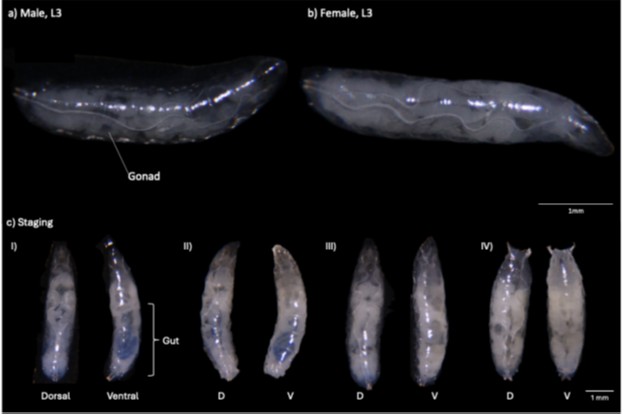
We thank the reviewer for this suggestion. We have now added reference images illustrating the WL3 and WPP stages for w1118 in the Appendix as suggested (Author response image 1). We have analysed the larvae-pupae transition in both sexes of miR-277-KO and miR-34-KO mutants, directly comparing them with stage-matched w1118 controls, and we did not observe any detectable differences in larval morphology, pupal morphology, anterior spiracle eversion, gut-purge status, or timing of pupariation between the mutants and controls.
Author response image 1.
(3) Biological validation of the identified sexually dimorphic genes in vivo will be necessary for the support of this work.
We have now included additional quantitative RT-PCR validation of key insect hormone pathway genes in male and female WL3 and WPP stages of wild-type, miR-277-KO, and miR-34-KO flies (Figure 5). These additional analyses confirm sex-specific expression differences in both wild-type and mutant backgrounds, supporting dimorphic endocrine regulation. The main text are also revised and shown as follows:
Lines 276-287 - “To understand how different hormones such as juvenile hormone (Belgacem and Martin, 2002) involved during metamorphosis in both sexes, we selected and tested a selection of candidates from both sesquiterpenoid and ecdysteroid hormone pathways, and found JHAMT, Spok, CG40486, and CG10962 showed differential expression during transition from WL3 to WPP in mutant and/or wild-type fly lines in the transcriptome analyses (Supplementary Figure 16). To further examine these RNA-seq patterns, we performed RT-PCR on nine candidate genes from WL3 to WPP in WT and mutant fly lines. In the larval-pupal transition comparison, CG40486 was consistent down-regulated in WT males and females in both RNA-seq and RT-qPCR datasets. In the mutant-vs-WT comparison, CG9360 showed concordant downregulation in miR-277-KO WPP females. Other tested genes showed more condition-specific patterns matching between the transcriptomic and RT-qPCR analyses. Further validation and mechanistic studies will be required to define how these hormone-pathway changes contribute to the larval–pupal transition.”
Reviewer #3 (Public Review):
Summary:
The authors show convincingly the complexity of gene up- and down-regulation at the outset of metamorphosis and identify substantial differences between the two sexes, even at this early time in development. The complexity of microRNA expression and the difference between the sexes are also nicely laid out. The functional significance of these differences, though, is harder to establish. The authors have focused on the roles of two families of developmental hormones, the ecdysteroids, and the juvenile hormones. The emergence of sex-specific differentiation of organs during metamorphosis is clearly downstream of the action of ecdysteroids and/or JH, but there is no evidence that the presence or lack of these hormones has any effect on the sexual identity of organ systems - i.e., that manipulations of JH or ecdysteroid result in either the masculinization or feminization of individuals or their organs. The precedence for the linkage of these hormones to sex determination is the 2002, Belgacem & Martin study, which describes the effects of JH on fly locomotion. These authors show that the number of stop/start bouts is sexually dimorphic, and removal of JH in males shifts their frequency into the female range while giving treated males exogenous JH moves it back. While this is referred to as a "feminization" of male behavior, this quantitative shift in frequency is not as compelling as would be a qualitative shift -- for example, the removal of JH causing males to show egg-laying behavior (a result that has never been seen). Also, these effects are in a fully mature system, rather than at the early metamorphic time examined in the present paper. In driving and coordination metamorphosis, JH and ecdysteroids are intimately involved in sexual differentiation, but I know of no compelling evidence that they play a role in sex determination.
We thank the reviewer for the positive and constructive comments. We agree that our findings advanced on the sexual differences in gene regulation during metamorphosis rather than establishing direct evidence of juvenile hormone (JH) or ecdysteroids in influencing insect sex determination. In addition to rephrasing at various parts of the main text (see below), we have also carried out additional experiments in obtaining and analysing transcriptomes of late larvae and early pupae of both sexes in the wild-type of another species (Drosophila virilis), which showed similar KEGG pathways enriched. The revised parts are shown as follows:
Lines 289-303 – Results section
“Metamorphosis between different sexes in D. melanogaster and D. virilis
In D. melanogaster, the transition from WL3 to WPP shows increased carbohydrate/energy metabolism and ER protein processing, with decreased lipid metabolism, detoxification (cytochrome P450), and redox/cofactor pathways (folate, glutathione, retinol); while PM-LM exhibits broader negative enrichment in translational and RNA-processing machinery (ribosome, spliceosome, proteasome, RNA degradation, ubiquitin-mediated proteolysis) and in signaling modules (mTOR, MAPK, FoxO, Hippo), whereas PF-LF shifts are smaller and less coordinated by GSEA (Supplementary Figure S15). On the other hand in D. virilis, core features match that of D. melanogaster from WL3 to WPP, carbohydrate/energy metabolism and ER protein processing increase, while lipid, detoxification, and redox/cofactor pathways decrease. The sex bias is inverted relative to D. melanogaster as PM-LM shows limited up-regulation, whereas PF-LF displays broader, coordinated repression involving amino-acid metabolism (glycine/serine/threonine; cysteine/methionine), lysosome, ABC transporters, and longevity-related signaling, yielding the largest DEG set in females (Supplementary Figure S15). These data strongly suggested the sexual differences in gene regulation during metamorphosis in insects is not species-specific.”
Lines 354-361 – Methods section
“D. melanogaster w1118 and D. virilis were reared at 25 C, 45-50% relative humidity and 14:10 hour light-dark cycle. Sexes of wandering L3 larvae instar (WL3) were differentiated under the light dissecting microscope by the presence (male) or absence (female) of gonads (Supplementary Figure 1). Collections were restricted to larvae wandering out of food and ready for pupariation. WPP were defined as immobile puparia with everted spiracles and a uniform white cuticle that were no longer moving even with external stimuli. A total of three biological replicates (Whole-body of 1 individual was served as per biological replicate) per stage and sex were obtained for D. melanogaster and D. virilis.”
While the summary of the effects or removal of specific microRNAs on the components of the biosynthetic pathway for the JHs and ecdysteroids (Figure 2E ,F) is quite compelling, I am concerned about the effects of the removal of mir-277 and mir-34 on the levels of both the JHs and 20E. My concern centers around the data from the control group (w[1118] animals in Figure 2D). These data are the first report of a marked sex difference in the titer of either JH or ecdysteroid at the start of metamorphosis in Drosophila. As expected, males show a 10-20 increase in levels of JH III, JHB3, and 20E between the L3 stage and the white puparium, but, surprisingly, the levels of these hormones in female L3 larvae are equal to or greater than that seen at pupariation! These data for females run counter to over 50 years of work on the effects of ecdysteroids in Drosophila!
We thank the reviewer and agree that the endocrine profiles of WL3 females require careful interpretation, given the late-L3 to WPP transition represents a highly dynamic window in which ecdysteroid and sesquiterpenoid titres can fluctuate over short developmental intervals. The revised text is shown as follows:
Lines 322-326 – “Given the WL3 to WPP transition represents a highly dynamic window in which ecdysteroid and sesquiterpenoid titres can fluctuate over short developmental intervals, future experiments on narrower early-/mid-/late-wandering timepoints will be required to understand the dynamics of the microRNAs and hormones.”
As far as I can gather from the paper, the L3 data were obtained using wandering larvae. This stage lasts for about 12 hours and ends with pupariation. Larvae from this period need to be used with caution for hormone studies. Levels of both JH and ecdysteroid are low as larvae leave the food but rapidly rise to their peak levels at the white puparium stage 12 hours later. To deal with the rapidly changing hormonal landscape through this period, the researchers have used physiological markers to track this progression. Initially, it was the sage of "puffing" of the giant salivary gland chromosomes, but, for bulk collection of staged larvae, larvae are fed on food containing a blue dye, and progression is tracked by the loss of blue coloring from the gut. I could not find if the authors had any criteria for selecting larvae during the wandering period. Male and female larvae grow to different sizes. Might this difference in growth be biased when larvae were selected during their wandering phase?
We thank the reviewer and agree that WL3 represents a hormonally dynamic 12-hour window. To minimise developmental heterogeneity, WL3 larvae were selected for larvae that had completely exited the food and climbing upwards of the fly vials. To ensure tight developmental synchrony, pupating individuals from the same batch were collected 4 hours later, allowing us to match WL3 and WPP animals from a single, developmentally coordinated cohort. This paired-batch collection tried to minimise variation arising from differences in timing of wandering onset. As for the differences between the body sizes of male and female larvae, we minimise size-dependent bias, hormone titres measurement were measured to ng hormone per gram of tissue in our study, which corrects for sex-specific differences in mass. The information is now included in the main text as follows:
Lines 276-287 – “To understand how different hormones such as juvenile hormone (Belgacem and Martin, 2002) involved during metamorphosis in both sexes, we selected and tested a selection of candidates from both sesquiterpenoid and ecdysteroid hormone pathways, and found JHAMT, Spok, CG40486, and CG10962 showed differential expression during transition from WL3 to WPP in mutant and/or wild-type fly lines in the transcriptome analyses (Supplementary Figure 16). To further examine these RNA-seq patterns, we performed RT-PCR on nine candidate genes from WL3 to WPP in WT and mutant fly lines. In the larval-pupal transition comparison, CG40486 was consistent down-regulated in WT males and females in both RNA-seq and RT-qPCR datasets. In the mutant-vs-WT comparison, CG9360 showed concordant downregulation in miR-277-KO WPP females. Other tested genes showed more condition-specific patterns matching between the transcriptomic and RT-qPCR analyses. Further validation and mechanistic studies will be required to define how these hormone-pathway changes contribute to the larval–pupal transition”.
The other hormone-related issue is the expression of Kr-h1 during larval stages and metamorphosis (Figure 1G). Kr-h1 is the main target of JH and Kr-h1 expression is often used as a proxy for the JH titer. The authors report that peak Kr-h1 expression occurs in the L3 (when the JH titer should be lowest!) and that it drops at wandering. This pattern is counter to that reported in the literature (e.g., FlyBase, ModEncode).
We have now revised the main text as follows:
Lines 204-207 – “It should be noticed that the analyses of this study was based on RNA taken from whole-body rather than individual tissue, and further experiments will be required to dissect the dynamics of Kr-h1 and different hormones both spatially and temporally.”
Besides dealing with the issues above, it would be useful in the background to establish the difference between "sex determination" and "sexual differentiation". I was surprised that the authors could deal with this subject without ever mentioning the sex determination pathway - e.g., double sex, transformer, etc.
In this study, one major focus was on the differences between sex during metamorphosis rather than the sex determination pathway. We have included the reviewer’s suggestion by adding the following information in the main text, shown as follows:
Lines 73-75 - “In insects, various genes related to sex determination or showing sexual dimorphic features have been revealed, including doublesex and transformer (Shukla and Nagaraju, 2010; Deng et al, 2025).”
Line 61: at the time that the authors are considering, -- the transition from larva to pupa - the main controller of ecdysone production in the prothoracicotropic hormone, not ecdysis triggering hormone.
Thanks for the reviewer’s suggestions. The main text is now written as follows:
Lines 44-47 - “In general, these three hormones work in a cooperative manner, where ecdysteroid 20-hydroxyecdysone controls the timing of ecdysis or molting, prothoracicotropic hormone and the insulin-like peptides act as regulators of ecdysteroid synthesis, and sesquiterpenoid hormones such as juvenile hormone (JH) regulates metamorphosis based on their titre levels.”
Line 71: it would be useful to name the main microRNAs that have been implicated in regulating insect metamorphosis.
The main text is now written as follows:
Lines 60-63 - “Previous studies have identified a number of microRNAs contributing to the regulation of insect metamorphosis such as let-7, miR-125, and miR-2 family (e.g. Caygill and Johnston 2008; Sokol et al 2008; Lozano et al 2015).”
For Figures 2E and F as well as 3C (and a number of supplementary figures), it would be useful to provide some quantitative representation of the level of activation or suppression (as done in Figure 3B) rather than just Red or Green
The trend of expression change of genes in selected pathways and detailed quantitative information can now be found in the supplementary file.



