
Peer review process
Revised: This Reviewed Preprint has been revised by the authors in response to the previous round of peer review; the eLife assessment and the public reviews have been updated where necessary by the editors and peer reviewers.
Read more about eLife’s peer review process.Editors
- Reviewing EditorJerry WorkmanStowers Institute for Medical Research, Kansas City, United States of America
- Senior EditorAdèle MarstonUniversity of Edinburgh, Edinburgh, United Kingdom
Reviewer #1 (Public review):
This work reports the finding of a dissociable human Mediator complex subunit, MED16, and its recruitment of transcription factors UBP1 and TFCP2 that form a trimeric complex regulating cellular gene transcription and inhibiting HIV promoter activity, thus promoting MED16-targeting inhibitors as HIV latency-reversing agents for anti-HIV therapy. In this revision, the authors provide new data indicating that MED16 action occurs in the holo-Mediator complex, not as a free form, for its functional association with UBP1 and TFCP2.
Summary:
Characterization of a dissociable Mediator subunit implicated in cellular pathways, particularly lung alveolar function, and HIV latency is conceptually interesting.
Strengths:
The strengths of this study are:
(1) Demonstration of MED16 dissociation from the core Mediator complex and formation of a subcomplex containing MED16, upstream-binding protein 1 (UBP1), and transcription factor cellular promoter 2 (TFCP2) by elegant biochemical fractionation and immunoblotting analysis.
(2) Defining nine N-terminal WD-40 repeats (WDRs) of MED16 as a Mediator-incorporating module and the C-terminal ⍺β-domain (157 amino acids) important for interaction with the UBP1-TFCP2 heterodimeric complex.
(3) Illustration of a weak hydrophobic interaction between MED16 and the Mediator core that could be disrupted by 1,6-hexanediol, but not by its 2,5-hexanediol isomer nor by high salt (500 mM NaCl) disruption.
(4) Classification of UBP1-upregulated cellular genes typically containing binding sites flanking the transcription start site (TSS) in contrast to UBP1-downregulated genes often containing a TSS-overlapping UBP1-binding site;
(5) Presenting evidence for Mediator complex-dissociated free MED16-repressed HIV promoter activity through functional association with UBP1 and showing bromodomain-containing protein 4 (BRD4) inhibitor JQ1 that potentially disrupts BRD4-inhibited HIV-1 transcription elongation could lead to reversal of HIV-1 latency.
Weaknesses:
(1) No clear demonstration that MED16-UBP1-TFCP2 indeed form a trimeric core subcomplex in regulating cellular gene transcription and HIV-1 promoter inhibition.
(2) No validation of transcriptomic datasets and pathways identified.
(3) Use of mostly artificial reporter gene constructs and non-HIV host cells (e.g., human 293T embryotic kidney cells, human HeLa cervical cancer cells, and mouse HT pancreatic cancer cells) for examining MED16/UBP1-regulated HIV transcription.
(4) Inconsistent use of 293T and HeLa cells in the characterization of dissociated MED16 interaction with UBP1 and TFCP2.
(5) In vitro transcription using immobilized DNA templates was not performed to a high standard thus failing to convincingly show MED16/UBP1-inhibited HIV-1 transcription preinitiation complex formation.
In this revision, the authors stipulate that the action of MED16-UBP1-TFCP2 is through the holo-Mediator complex, not as a free trimeric complex, since deletion of the N-terminal Mediator-incorporating module abolishes the functional effect of MED16 even though its interaction with UBP1-TFCP2 persists. The authors also show that a predicted UBP1-binding site is indeed present in the promoter region not overlapping with the TSS in each of the UBP1/MED16-upregulated respiratory gaseous exchange genes, such as surfactant protein A1 (SFTPA1), SFTPA2, SFTPB and SFTPC. To demonstrate UBP1/MED16-inhibited HIV transcription in a more biologically relevant system, the authors employ an HIV-1 latency model derived from CD4+ T cells. Additional experiments are also performed to address other issues raised by the reviewers.
Overall, the revised manuscript has been improved by addressing some of the reviewer's comments. Additional issues that came up while reading this revision are: a) The statement, "UBP1 (as known as LBP-1) and TFCP2 (as known as LSF) belong to a subfamily of the TFCP2/Grainyhead transcription factor family" (lines 63-64), is confusing and needs to be clarified. My understanding is that LBP-1 has four family members encoded by two different genes (see Yoon et al., MCB 14: 1776-1785, 1994): LBP-1a and LBP-1b are probably UBP1, and LBP-1c and LBP-1d are likely TFCP2/LSF. If this is correct, the authors should clearly indicate which specific isoforms are analyzed in their UBP1 and TFCP2 proteins and clarify some of the text descriptions seemingly contradictory to the earlier findings.
The use of mostly artificial reporter templates and transfection assays conducted mostly in 293T and HeLa cells remains concerning. It is also unclear why overexpression of MED16 only, but not MED23 or MED24, inhibits HIV promoter activity (Figure 5) if MED16 action is through the holo-Mediator complex and overexpression of MED16 may titrate out key cellular factors, thus diminishing their action through holo-Mediator. Results based on this squelching type of experiment relying on overexpression of a single component needs to be carefully interpreted.
Reviewer #2 (Public review):
Summary:
The article from Zheng et al. proposes that the Med16 subunit of Mediator can dissociate from the complex, associate with transcription factor UBP1, and contribute to activation or repression of specific sets of genes in human cells. The question is potentially interesting, and the possibility of a distinct role for Med16 in UBP1-dependent transcription could be of interest to the field. However, I have substantial concerns about the experimental design, reproducibility, and interpretation of the data. In particular, several of the central conclusions rely on single experiments, indirect assays, or perturbations that are expected to cause broader changes in Mediator structure and transcription. In my view, the current data therefore do not provide sufficient evidence to support several of the principal mechanistic conclusions of the manuscript.
Strengths:
The authors provide preliminary evidence that Med16 may exist in an altered or partially dissociated Mediator complex and may be associated with specific transcriptional effects. The use of biochemical assays, reporter assays, RNA-seq, and Med16 knockout/rescue experiments provides several potentially useful approaches to investigating this question. In particular, the Med16 knockout/rescue RNA-seq experiment described in the rebuttal is a potentially informative experiment. Making the underlying IP-MS abundance and peptide-count data available is also an improvement over the previous version.
However, important limitations of these experiments need to be addressed before the proposed mechanism can be established.
Comments on the revised version:
In the previous review, I raised concerns that the evidence for a specific UBP1-Med16 interaction and for a mechanistic role of this interaction was limited. The revised manuscript does not resolve these concerns. In particular, whether UBP1 interacts specifically with Med16, whether this interaction is required for activation of UBP1 target genes, and whether UBP1 represses HIV-1 transcription through Med16 remain unresolved.
A stronger case for a specific UBP1-Med16 interaction would require more direct biochemical, structural, or biophysical evidence. The current experiments do not distinguish sufficiently between a specific Med16-dependent mechanism and broader consequences of perturbing Mediator structure or transcription.
The revision also does not include the additional biological replicates needed to assess the reproducibility of several foundational experiments. Consequently, a number of the central conclusions remain based on single experiments, which substantially limits the strength of the conclusions that can be drawn.
Major concerns:
Figure 1:
All of the data in Figure 1 are from single experiments without biological replicates. Because Figure 1 provides an important foundation for the proposed mechanism, this limits confidence in the reproducibility of the observations. Western blot experiments can show substantial experiment-to-experiment variability, and such variability appears relevant to several of the observations presented here.
A key experiment is the western blot analysis of the gel-filtration experiment in Figure 1D. Rather than migrating with Mediator, the majority of the detected Med16 appears to elute at a lower molecular weight. The extract was maintained at super-physiological salt concentrations from preparation through column loading and elution, which could potentially contribute to this result. Other possibilities include experiment-to-experiment variation or detection of a cross-reacting protein by the Med16 antibody. The latter possibility warrants consideration because the bands detected in fractions 28-31 appear to migrate at a different molecular weight from the Med16 bands in fractions 16-19. Additional replicated experiments and appropriate controls would be needed to establish that the lower-molecular-weight signal represents Med16 in a distinct complex.
Figure 2:
The data in Figure 2 are also based on single experiments without biological replicates. The biochemical experiments use protein fragments that do not occur as such in the native protein. Such experiments can be useful for mapping possible protein-protein interaction surfaces, but they can also generate false-positive or false-negative interactions depending on the experimental configuration.
Some of the results raise additional questions about the proposed interaction model. For example, the observations involving TFCP2 in panel B and Med1 and Med23 in panel F appear difficult to reconcile with a simple model in which UBP1 interacts with Mediator through Med16. Panel 2F is particularly relevant because Med1 and Med23 are not detected despite the proposed model involving UBP1 interaction with Mediator. These observations should be addressed when interpreting the specificity and mechanism of the proposed UBP1-Med16 interaction.
Figure 3:
The reporter assays provide potentially useful preliminary evidence for factors that influence transcription. However, the relatively long experimental timeframes (36-48 hours after transfection), use of artificial promoters, and overexpression of proteins introduce important limitations.
The data are consistent with the authors' model, but they are also consistent with a simpler explanation in which Med16 depletion broadly affects Mediator structure and function. Previous work from the Gross, Robert, and Roeder laboratories has shown that loss of Med16 can result in dissociation of Med23, Med24, and Med25 from Mediator. Importantly, the authors' own data in the rebuttal document appear to show similar effects following Med16 loss. These observations are important because Med23, Med24, and Med25 provide major interfaces for DNA-binding transcription factors, and Med16, Med23, Med24, and Med25 together represent a substantial fraction of the molecular mass of Mediator. The functional properties and stability of the resulting Mediator complex therefore need to be considered when interpreting Med16 depletion experiments.
The authors' data also suggest broader transcriptional effects: Med16 knockout causes loss of basal transcription in panel 3E. Conversely, overexpression of Med16 or Med16 fragments can cause broad defects in transcriptional activation, including through squelching. The relatively modest effects observed despite substantial increases in protein abundance are therefore also consistent with non-specific consequences of overexpression. These alternative explanations need to be considered when interpreting the reporter assays.
Figure 4:
RNA-seq is an appropriate approach for investigating possible effects of Med16 on gene expression. However, the experimental design has important limitations because Med16 depletion is expected to affect the composition of Mediator, including loss of Med23, Med24, and Med25. The knockdown also occurs over an extended timeframe (>24 hours), making it difficult to distinguish direct effects of Med16 from secondary effects arising from altered Mediator structure or broader changes in transcription.
The RNA-seq results could therefore reflect Med16-specific functions, but they are also consistent with a model in which loss of Med16 compromises Mediator structure and function and produces downstream gene-expression changes.
The presentation of the RNA-seq data in Figure 4E is also difficult to interpret. The volcano plot shows UBP1 overexpression versus the shCTRL vector, whereas the comparison central to the authors' interpretation concerns the effects of shMed16. Showing the volcano plot for the shMed16 comparison would allow readers to assess the statistical significance and magnitude of the relevant gene-expression changes directly. Instead, the authors superimpose "x" symbols for genes that "failed to be activated by UBP1 in the shMed16 cell line." Because the underlying Med16 comparison is not displayed, the p-values and fold changes for the comparison of interest cannot be evaluated. Moreover, positioning the "x" symbols according to the coordinates of the UBP1-overexpression volcano plot does not appear to represent their corresponding positions in a volcano plot of the Med16 comparison. This makes the figure difficult to interpret and could give a misleading visual impression of the underlying data.
Finally, the RNA-seq data are not spike-in normalized. This is potentially important because Med16 depletion is expected to cause broad effects on transcription and could result in an overall reduction in transcription across the genome. Appropriate normalization is therefore important for distinguishing gene-specific effects from global changes. I also did not identify information in the manuscript indicating where the RNA-seq data have been deposited in a public repository.
Figure 5:
The reporter assays in Figure 5 are subject to similar concerns. The authors' model may explain the observations, but the data are also consistent with broader disruption of Mediator structure and function following Med16 depletion, or with more general transcriptional effects caused by overexpression of Med16 or Med16 fragments.
A separate concern is the proposed mechanism for HIV-1 repression. The authors propose that UBP1 interacts with Med16/Mediator and that overexpression of Med16 enhances Mediator recruitment, although Mediator recruitment is not directly measured. The proposed increase in Mediator recruitment would, in isolation, be expected to promote transcription rather than repression. The authors instead invoke a UBP1-dependent mechanism to explain the repression. However, a simpler explanation is that overexpression of Med16 or selected Med16 fragments perturbs normal transcription through mechanisms including squelching. The authors themselves invoke a Med16 squelching model to explain reduced transcription at the UBP1 reporter in Figure 3G-I. The possibility that the same mechanism contributes to the results in Figure 5 should therefore be addressed.
Finally, a central component of the proposed model is recruitment of Mediator by UBP1 through Med16, but neither UBP1 occupancy nor Mediator occupancy at the relevant promoters is directly measured. These experiments would provide important evidence for the proposed mechanism. There is also no legend for Figure 5G,H, and the legend for Figure 5C,D appears to be incorrect.
Figure 6:
The title of Figure 6 states that "The position of the UBP1 binding site relative to the TSS dictates whether MED16-UBP1 activates or represses transcription." The data presented do not establish this conclusion.
Only one alternative location of the UBP1 binding site is tested, and the analysis in panel D compares only the 50 most up- or down-regulated genes in UBP1-overexpression experiments. The correlations shown are relatively weak, and a rigorous statistical analysis is not presented.
The authors cite Duttke et al. (Nature 2024) in support of the concept that transcription-factor position can influence regulatory activity. That study used genome-wide mapping of native TSSs, MPRA analysis of approximately 2,000 promoter variants, and targeted follow-up experiments. By contrast, Figure 6 relies on an artificial reporter and a single change in UBP1 binding-site position. The current data therefore support the possibility that binding-site position influences the observed reporter activity, but they do not establish the broader conclusion that the position of the UBP1 binding site relative to the TSS dictates whether Med16-UBP1 activates or represses transcription.
Figure 7:
The title of Figure 7 states that Med16 "interferes with HIV-1 PIC assembly," but the data presented do not establish this mechanism.
First, the proposed mechanism is described as acting through interaction with UBP1, but UBP1 is not measured in this experiment.
Second, Med16 knockout or overexpression has relatively modest effects on the estimated levels of bound PIC components, and these changes do not consistently correspond to the transcriptional changes shown in panel C. For example, in the WT versus Med16 KO comparison, there is no apparent difference in the levels of Pol II, Med1, IIB, or IIH based on the western blot data. The reduction in Med16 and Med23 is expected following Med16 knockout and therefore does not by itself establish altered PIC assembly. In the WT versus Med16 overexpression comparison, panel C suggests a meaningful reduction in transcription, whereas the changes in PIC-associated proteins do not follow the direction predicted by the proposed model: Pol II and IIB increase with Med16 overexpression, while Med1, Med23, Med16, and IIH appear similar.
Third, Cdk8 and Med12 are not PIC components. They are subunits of the CDK8 module, which can inhibit PIC formation. Even under a simplified interpretation in which reduced Cdk8 and Med12 levels were relevant to PIC formation, the direction of these changes is opposite to that expected from the "relative mRNA level" plots in panel C. Reduced levels of these proteins in the Med16 overexpression condition would, under this interpretation, be expected to promote rather than reduce transcription.
Finally, this is a single-replicate experiment, and quantitative comparisons are made across immobilized-template pulldowns using different nuclear extracts (WT, Med16 KO, and Med16 OE). Differences in protein concentration between extracts, differences in pulldown efficiency, and experiment-to-experiment variation could all contribute to the observed differences. Replicated experiments with appropriate normalization would therefore be needed to support the proposed PIC mechanism.
Figure 8:
Panel F presents a model that, in my view, goes beyond what is supported by the experimental data. In particular, there is currently insufficient evidence to establish the proposed Med16-UBP1-TFCP2 complex as a distinct functional complex.
The model also depicts the Mediator CDK8 module bound at promoters together with Pol II. The manuscript does not provide evidence supporting this arrangement, and there is substantial prior evidence that the CDK8 module can inhibit or prevent PIC formation. This aspect of the model therefore requires either experimental support or clarification.
Finally, the model depicts a separate Med16-UBP1-TFCP2 complex. This appears inconsistent with the authors' statements in the rebuttal that they did not intend to propose such an unsupported isolated complex. If this is not the intended model, Figure 8 should be revised to avoid conveying that interpretation.
Figure S1: IP-MS analysis:
The IP-MS data raise substantial concerns regarding the specificity of the proposed UBP1-Med16 interaction.
The authors state that they "have now reanalyzed the IP-MS data using a more precise quantification approach." However, the filtering procedure described in the Methods is difficult to evaluate and requires substantially more justification. The procedure includes multiple sequential criteria: detection in at least two of three replicates of the target bait; a minimum unique peptide count; detection in at least one additional Mediator bait; exclusion of Mediator/Pol II subunits, ribosomal proteins, histones, keratins, and common contaminants; and annotation with transcriptional or DNA-binding functions using GO terms. The rationale for this particular combination of filters and its impact on the candidate list should be explained and justified.
The resulting "bait-preferential score" is also difficult to assess. The reported mean_target and mean_other values are frequently negative, with some positive values, and the method appears to derive a positive score from the difference between these values after taking an absolute value. The biological and statistical interpretation of this score is therefore unclear. The reported scores are generally clustered around relatively narrow values, and the analysis does not appear to provide the statistical framework normally used to assess enrichment and reproducibility in IP-MS experiments.
Importantly, the candidate proteins identified by this approach do not appear to overlap with proteins identified in other published Med16 IP-MS analyses in human cell lines, including HCT116, MCF7, HEK293T, and HeLa. I was unable to identify a protein among the 37 Med16 candidates that was consistently observed in the cited resources, including BioPlex (Huttlin et al., Cell 2021) and Malovannaya et al. (Cell 2011). This lack of concordance raises questions about the specificity and physiological relevance of the proposed Med16-associated proteins and, in particular, about the evidence for a specific UBP1-Med16 interaction.
The peptide counts for UBP1 also illustrate the limitation of the current analysis. The total UBP1 peptide counts across the Cdk8, Med1, and Med16 IP-MS experiments are 12, 10, and 14, respectively, yielding bait-preferential scores of 1.07, 0.76, and 1.85. Given these relatively small differences and the absence of statistical significance measures, it is difficult to determine whether these observations provide evidence for a specific biological interaction.
The authors have now made the raw abundance values and peptide counts available as supplementary tables, which is useful. However, standard statistical measures for evaluating MS data, including FDR, p-values, and adjusted p-values, are not provided, nor is sufficient information given about how the reported scores were calculated. These additions would be important for readers to assess the robustness of the conclusions.
Concerns about the rebuttal document:
Several statements in the rebuttal appear inconsistent with the data shown in the revised manuscript or with the interpretation of the authors' own experiments. I highlight specific examples below.
(1) The proposed Med16-UBP1 complex:
I previously raised concerns that the manuscript overstated the evidence for a separate Med16 complex associated with UBP1 and lacked rigorous biochemical characterization of such a complex. The authors responded that the wording of the original manuscript may have created this interpretation and stated that this was not the mechanistic model they intended to convey. However, Figure 8 of the revised manuscript still depicts a separate Med16-UBP1-TFCP2 complex. The figure therefore appears to retain the interpretation that the authors stated they did not intend to convey. This should be clarified.
(2) Replication and experimental rigor:
I previously raised concerns about the absence of replication and the limitations of the experimental approaches used to infer roles for Med16 and UBP1. These concerns remain because no additional replicate experiments were performed for several of the key experiments. As a result, important conclusions continue to depend on single western blots, including foundational experiments in Figures 1, 2, and 7.
Examples include the variable quantification in Figure 1A; the absence of detectable Med23 and Med1 in the UBP1 IP in Figure 2F despite the proposed interaction between UBP1 and Mediator; and the variable association of TFCP2 with Flag-MED16 fragments in Figure 2B, including lanes 7-9. Replication would be important for determining whether these observations are reproducible.
(3) IP-MS analysis:
The revised IP-MS analysis provides additional information compared with the previous version, but important concerns remain regarding the processing and interpretation of the data. The "bait-preferential score" approach is not sufficiently justified, and standard measures such as p-values, adjusted p-values, or FDR are not provided.
The analysis also identifies no proteins among the 37 Med16 candidates that appear to be consistently observed in other published Med16 IP-MS analyses in human cell lines. This is important when assessing the specificity of the proposed interaction.
The peptide counts for UBP1 are similarly low and do not show a clear Med16-specific enrichment: 12 peptides in the Cdk8 IP, 10 in the Med1 IP, and 14 in the Med16 IP, corresponding to scores of 1.07, 0.76, and 1.85, respectively. These observations do not by themselves provide strong evidence for a specific UBP1-Med16 interaction.
In addition, the Med16 IP experiments identify approximately 800 proteins per replicate. This broad set of associated proteins highlights the importance of appropriate controls and statistical analysis when interpreting a co-IP as evidence for a specific protein-protein interaction.
(4) Interpretation of the 1,6-HD experiments:
I previously raised concerns that the IP-western data in Figure 1 did not establish a Med16-specific effect. The rebuttal states that "MED16 exhibits a uniquely pronounced dissociation by 1,6-HD that is consistent across three independent IPs using anti-CDK8, anti-MED1, and anti-MED12 antibodies." However, the quantitative data shown do not clearly support this interpretation.
In the CDK8 IP, CDK8 and MED23 appear to be lost to a similar extent as Med16. In the Med1 IP, Med12 shows a similar loss to Med16, with Med23 also showing a comparable trend. In the Med12 IP, CDK8 and Med23 show changes comparable to Med16. There are also unexplained increases in some Mediator subunits with increasing 1,6-HD concentrations. For example, Med12 and Med1 are variable across the 1,6-HD conditions, and Med1 and Med24 increase at higher concentrations. Similar trends are present in the CDK8 IP.
The Figure 1B data raise related concerns. Several Mediator subunits show substantial increases following the 2,5-HD condition compared with 1,6-HD, in some cases greater than the change observed for Med16. Thus, the available data do not clearly establish that Med16 exhibits a uniquely pronounced dissociation from Mediator.
(5) Evidence for the proposed mechanism:
I previously suggested that more rigorous approaches would be needed to distinguish a specific Med16-dependent mechanism from broader effects of Med16 perturbation. The rebuttal instead emphasizes that the authors' conclusion that the UBP1-TFCP2 heterodimer acts through Med16 is supported by multiple independent lines of evidence.
However, the different experiments do not independently establish the same mechanistic conclusion. Many rely on long experimental timeframes, overexpression, reporter assays, limited controls, or lack of biological replication. These approaches can provide useful preliminary evidence for effects on transcription but do not establish that a specific UBP1-Med16 interaction is required for UBP1 target-gene expression.
Additional approaches that directly test the proposed interaction and its requirement for transcriptional regulation would therefore be needed to establish this mechanism.
(6) Med16 rescue experiment:
The authors performed an RNA-seq comparison of WT, Med16 KO, and Med16 KO cells rescued by re-expression of WT Med16. This is a potentially informative experiment, and I appreciate that it was performed.
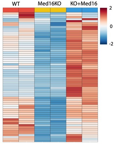
However, the heatmap shown in the rebuttal does not appear to demonstrate a clear rescue of UBP1 target-gene expression. The figure contains approximately 60 genes but provides no legend beyond "Gene profiling of MED16 KO and rescue effect." The authors describe the result as showing that re-expression of Med16 can largely restore expression of UBP1 target genes, but this conclusion is not readily apparent from the figure as presented. More information about the analysis, normalization, and comparisons would be needed to evaluate the claimed rescue. It would also be useful to include the relevant analysis in the revised manuscript rather than only in the rebuttal.
(7) Consequences of Med16 depletion:
A continuing concern is that the manuscript does not adequately account for the broader consequences of Med16 depletion or knockout. Loss of Med16 is known to cause dissociation of other Mediator subunits, including Med23, Med24, and Med25. The authors acknowledge this in the rebuttal and state that their new Co-IP and IP-MS experiments confirm partial dissociation of these subunits following Med16 knockout. They also acknowledge that this structural perturbation must be considered when interpreting phenotypes from Med16 depletion and that long-term knockdown or knockout approaches are susceptible to secondary effects and cellular adaptation.
These are important caveats, but they are not adequately incorporated into the interpretation of the revised manuscript, and the new data demonstrating loss of Med23, Med24, and Med25 are not shown in the article.
This issue is particularly important for interpreting the transcriptional experiments. Loss of Med16 is expected to disrupt Mediator more broadly, including loss of Med23, Med24, and Med25, which provide important transcription-factor interaction surfaces. These changes could produce widespread transcriptional effects independently of a specific UBP1-Med16 mechanism. Given that the authors' own experiments now provide evidence for this structural perturbation, the possibility of indirect effects and altered Mediator function should be explicitly incorporated into the experimental interpretation.
Overall assessment:
The manuscript addresses an interesting question and presents several potentially useful observations concerning Med16, Mediator, and UBP1. However, the central mechanistic conclusions are currently not supported by sufficiently rigorous or independent evidence. The principal concerns are the extensive reliance on single-replicate experiments; the use of overexpression, artificial reporters, and long depletion timeframes; the absence of direct measurements of UBP1 and Mediator occupancy; the broad structural consequences of Med16 depletion; and the limited statistical support for the IP-MS analysis.
Taken together, the current data do not distinguish adequately between the proposed specific UBP1-Med16 mechanism and alternative explanations involving broader disruption of Mediator structure or transcription. Addressing this distinction is important because it affects the interpretation of much of the manuscript, including the RNA-seq and reporter-assay results and the proposed HIV-1 mechanism.
The authors have generated several useful datasets, including the Med16 knockout/rescue experiment and additional IP-MS information, but the manuscript would need to incorporate the associated caveats and provide stronger experimental evidence before the proposed mechanistic model can be established.
Author response:
The following is the authors’ response to the original reviews.
Public Reviews:
Reviewer #1 (Public review):
Summary:
Characterization of a dissociable Mediator subunit implicated in cellular pathways, particularly lung alveolar function, and HIV latency is conceptually interesting.
Strengths:
The strengths of this study are:
(1) Demonstration of MED16 dissociation from the core Mediator complex and formation of a subcomplex containing MED16, upstream-binding protein 1 (UBP1), and transcription factor cellular promoter 2 (TFCP2) by elegant biochemical fractionation and immunoblotting analysis.
(2) Defining nine N-terminal WD-40 repeats (WDRs) of MED16 as a Mediator-incorporating module and the C-terminal ⍺β-domain (157 amino acids) important for interaction with the UBP1-TFCP2 heterodimeric complex.
(3) Illustration of a weak hydrophobic interaction between MED16 and the Mediator core that could be disrupted by 1,6-hexanediol, but not by its 2,5-hexanediol isomer nor by high salt (500 mM NaCl) disruption.
(4) Classification of UBP1-upregulated cellular genes typically containing binding sites flanking the transcription start site (TSS) in contrast to UBP1-downregulated genes often containing a TSS-overlapping UBP1-binding site
(5) Presenting evidence for Mediator complex-dissociated free MED16-repressed HIV promoter activity through functional association with UBP1 and showing bromodomain-containing protein 4 (BRD4) inhibitor JQ1 that potentially disrupts BRD4-inhibited HIV-1 transcription elongation could lead to reversal of HIV-1 latency.
Weaknesses:
Nevertheless, foreseeable weaknesses include:
(1) No clear demonstration of MED16-UBP1-TFCP2 indeed forming a trimeric core subcomplex in regulating cellular gene transcription and HIV-1 promoter inhibition
We appreciate this insightful comment from the reviewer. The physical interaction between MED16 and the UBP1–TFCP2 heterodimer was uncovered serendipitously via biochemical fractionation. Specifically, UBP1 and TFCP2 were identified in gel-filtration fractions containing free MED16 that eluted separately from the intact core Mediator complex (Figures 1D–1F). This biochemical observation suggested to us a potential functional linkage between MED16 and these two transcription factors. Subsequently, our functional assays further clarified this regulatory relationship: UBP1 and TFCP2 do interact with MED16 that is required to be fully assembled into Mediator complexes to exert transcriptional control. Truncation of MED16’s N-terminal WD40-repeat (WDR) domain (MED16ΔWDR) eliminates MED16 integration into the Mediator complex. Notably, this MED16ΔWDR mutant still retains an intact C-terminal αβ-domain capable of binding UBP1 yet completely loses the capacity to support UBP1-dependent transcription. Collectively, these data demonstrate that UBP1–TFCP2 engage MED16 as a component of the complete Mediator complex to govern target gene expression in cells.
We have updated the abstract and full manuscript to present this model more clearly and coherently throughout the text.
(2) No validation of transcriptomic datasets and pathways identified.
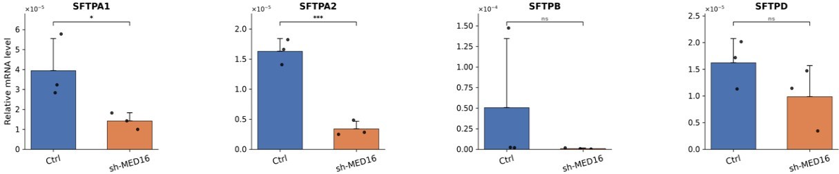
We thank the reviewer for this comment. Since our RNA-seq analysis identified SFTP family genes (SFTPA1, SFTPA2, SFTPB, SFTPD) as UBP1-MED16 co-regulated targets, and these are lung-specific surfactant protein genes, we have now performed qRT-PCR validation in the A549 human lung adenocarcinoma cell line. The results confirm that knockdown of MED16 largely attenuates the expression of these SFTP genes (see Author response image 1) and other UBP1 target genes identified in our RNA-seq dataset.
Author response image 1.
qRT-PCR validation of SFTP gene family expression (SFTPA1/A2/B/D) in A549 cells
(3) Use of mostly artificial reporter gene constructs and non-HIV host cells (e.g., human 293T embryonic kidney cells, human HeLa cervical cancer cells, and mouse HT pancreatic cancer cells) for examining MED16/UBP1-regulated HIV transcription.
To overcome the limitations of artificial reporter systems, we have managed to utilize multiple complementary approaches beyond reporter assays, including: (i) RNA-seq analysis of endogenous gene regulation, (ii) biochemical co-IP and gel filtration experiments, (iii) immobilized template transcription assays that directly measure PIC assembly and nascent RNA production, and (iv) validation in the J-Lat 10.6 CD4+ T cell line harboring an integrated latent HIV-1 provirus, where MED16 overexpression suppresses HIV-1 reactivation.
Unfortunately, the direct infection experiments with replication-competent HIV-1 were not feasible as our institution does not have the biosafety level 3 (BSL-3) containment facilities.
(4) Inconsistent use of 293T and HeLa cells in the characterization of dissociated MED16 interaction with UBP1 and TFCP2.
For biochemical characterization in this study, HEK293T cells were primarily utilized for overexpression-based co-immunoprecipitation (Co-IP) and domain-mapping assays owing to their superior transfection efficiency. In contrast, HeLa cells were employed for gel-filtration chromatography and endogenous Co-IP, as fractionation of HeLa nuclear extracts represents a well-established workflow for purifying native protein complexes. Critically, the physical interaction between MED16 and the UBP1–TFCP2 heterodimer was consistently detected in both HEK293T whole-cell lysates and HeLa nuclear extracts across all assays.
(5) In vitro transcription using immobilized DNA templates was not performed to a high standard, thus failing to convincingly show MED16/UBP1-inhibited HIV-1 transcription preinitiation complex formation.
We appreciate the reviewer’s careful assessment of the immobilized template assay. While we recognize the technical challenges inherent in this approach, we want to emphasize that the proposed model—that MED16-UBP1 inhibits HIV-1 transcription at the level of PIC assembly—does not rest solely on this single experiment.
Multiple independent lines of evidence converge on this model:
(1) The MED16-UBP1 inhibition depends on the UBP1 binding site overlapping the TSS: a chimeric reporter with the UBP1 binding site placed at the TSS is inhibited by MED16, whereas placing it upstream does not show inhibition, demonstrating the UBP1 binding site-specific repression (Fig 6A-C).
(2) MED16 overexpression inhibits HIV-1 reporter activity in a manner depending on interaction with UBP1. Deletion of the MED16 αβ-domain (MED16Δαβ), which abolishes MED16-UBP1 interaction, completely abrogates the inhibitory effect (Fig 5E-G).
(3) The MED16-UBP1 inhibition of HIV is independent of TAR-mediated elongation (Fig S2B-C) and HDAC-mediated deacetylation (Fig S2A), consistent with an inhibition mechanism operating at the initiation stage rather than at the post-initiation steps.
(4) Upon MED16 overexpression, the immobilized template assay shows reduced CDK8 binding and Pol II pSer5 signals, along with TFIIB retention, providing mechanistic evidence that is consistent with the aforementioned functional observations.
Thus, the model is supported by convergent evidence from reporter assays, domain-mapping experiments, pharmacological inhibition, and in vitro biochemical reconstitution. In the revised manuscript, we have further clarified how these complementary approaches collectively support the PIC inhibition model.
Reviewer #2 (Public review):
Summary:
The article from Zheng et al. proposes an interesting hypothesis that the Med16 subunit of Mediator detaches from the complex, associates with transcription factor UBP1, and this complex activates or represses specific sets of genes in human cells. Despite my excitement upon reading the abstract, I was concerned by the lack of rigor in the experimental design. The only statement in the abstract that has some experimental support is the finding that Med16 dissociates from the Mediator and forms a subcomplex, but the data shown remain incomplete.
We would like to first address a core conceptual point that we believe underpins several of the reviewer’s concerns.
We respectfully disagreed that the reviewer summarized our proposed model as a pathway where “Med16 detaches from the complex, associates with transcription factor UBP1, and this complex activates or represses specific sets of genes.” We acknowledge that phrasing in our original manuscript may have created this misleading interpretation, yet this is not the mechanistic model we intend to convey. We identified the MED16–UBP1–TFCP2 interaction serendipitously during biochemical fractionation: UBP1 and TFCP2 co-eluted with free MED16 in gel-filtration fractions that were fully separated from the intact core Mediator complex. While this biochemical result guided our subsequent functional investigation into MED16–UBP1–TFCP2 crosstalk, we never intend to propose that a standalone MED16–UBP1–TFCP2 subcomplex can drive transcriptional activation or repression independently of the intact Mediator complex.
Most importantly, our functional experiments demonstrate that UBP1 and TFCP2 require MED16 as an integral part of the Mediator complex to regulate transcription as shown by domain mapping experiments. Specifically, MED16 uses its N-terminal WDR domain to integrate into the Mediator and its C-terminal αβ-domain to bind UBP1/TFCP2. Deletion of either WDR or αβ-domain is functionally inert in both cellular and HIV-1 reporter assays. Thus, the MED16-UBP1 interaction is functionally relevant because it links these transcription factors to the Mediator complex, not because it forms an autonomous regulatory complex. In another word, UBP1–TFCP2 target MED16 to recruit the whole Mediator complex for governing gene expression in cells.
We have comprehensively revised the full manuscript to sharply distinguish our initial biochemical observation from the validated functional model. As all experimental approaches carry inherent limitations, we tried our best to validate every major conclusion using multiple orthogonal assays and to draw mechanistic interpretations when results from distinct experimental systems do converge. Our detailed replies to each specific concern from the reviewer are provided below.
Strengths:
The authors have preliminary evidence that a stable Med16 complex may exist and that it may regulate specific sets of genes.
Weaknesses:
The experiments are poorly designed and can only infer possible roles for Med16 or UBP1 at this point. Furthermore, the data are often of poor quality and lack replication and quantitation. In other cases, key data such as MS results aren't even shown. Instead, we are given a curated list of only about 6 proteins (Figure S1), a subset of which the authors chose to pursue with follow-up experiments. This is not the expected level of scientific process.
As clarified in our Summary Response above, we thought this reviewer may have misinterpreted our data and model. Overall, our work demonstrates that MED16 acts as a molecular bridge, linking UBP1/TFCP2 to the Mediator complex, not that three proteins form a free-standing subcomplex, and we have extensively revised the manuscript to clarify this.
Here we also want to address the specific concerns about data completeness and rigor. The full mass spectrometry datasets were not clearly presented in the original submission. We have now reanalyzed the IP-MS data using a more precise quantification approach. Rather than scoring proteins by simple presence or absence, we calculated a bait-preferential score for each protein by comparing its normalized abundance in the MED16 IP to that in the other three Mediator bait IPs (MED1, MED12, CDK8). This score reflects the degree to which a protein is preferentially enriched by MED16 relative to other Mediator subunits. The bait-specific candidates identified by this analysis are presented in Fig S1B, and the complete list of MED16-specific candidates is now included. In addition, the raw abundance values and peptide counts for all identified proteins have been compiled and uploaded as supplementary tables. A detailed description of the normalization and scoring procedure is provided in the Methods section
Furthermore, we have added quantification analysis for key Western blot results throughout the revised figures.
(1) The data supporting the Med16 dissociation and co-association with UBP1 are incomplete and not convincing at this stage. According to the Methods and text, the gel filtration column was run with "un-dialyzed HeLa cell nuclear extract" and eluted in 300mM KCl buffer. The extracts were generated with the Dignam/Roeder method according to the text. Undialyzed, that means the extract would be between 0.4 - 0.5M NaCl. Under these high salt conditions (not physiological), it's possible and even plausible that Mediator subunits could separate over time. This caveat is not mentioned or controlled for by the authors. Because a putative Med16 subcomplex is a foundational point of the article, this is concerning.
The data are incomplete because a potential Med16 complex is not defined biochemically. The current state suggests a smaller Med16-containing complex that may also contain UBP1 and other factors, but its composition is not determined. This is important because if you're going to conclude a new and biologically relevant Med16 complex, which is a point of the article, then readers will expect you to do that.
We appreciate the reviewer’s thorough feedback and address the two core concerns separately below.
(1) Undialyzed nuclear extracts & high-salt conditions
Published work confirms Mediator undergoes massive precipitation (~70% loss) upon dialysis to low-salt 100 mM KCl buffer (1), justifying our use of undialyzed HeLa nuclear extracts for biochemical assays. Prior study also demonstrated intact Mediator remains stable in up to 1 M KCl (1). Therefore, MED16 dissociation seen at 300 mM KCl arises from its unique biochemical property, rather than non-specific high-salt damage.
(2) Biochemical interpretation of MED16-containing fractions
We have revised ambiguous original wording to clarify our model: we do not intend to propose an autonomous MED16–UBP1–TFCP2 subcomplex functions independently in cells. Gel filtration only captures a biochemical state where MED16 co-elutes with UBP1/TFCP2 separate from core Mediator, which merely inspired our functional follow-up. Three lines of evidence prove UBP1/TFCP2 require fully assembled Mediator to function:
(i) MED16’s N-terminal WDR domain mediates Mediator integration, while its C-terminal αβ-domain binds UBP1; both interactions can occur simultaneously (Fig 2B).
(ii) MED16Δαβ incorporates into Mediator but loses UBP1 binding, showing no activity in UBP1-driven and HIV-1 reporter assays (Fig 3G, 5G).
(iii) MED16ΔWDR retains UBP1 binding yet cannot assemble into Mediator, thus fails to repress HIV-1 transcription (Fig 5F).
In vitro MED16 separation only reflects its extractable biochemical behavior, not a functional standalone complex. Collectively, our data establish UBP1/TFCP2 recruit the entire Mediator complex via MED16. All relevant manuscript sections have been rewritten to distinguish this preliminary biochemical observation from our validated functional model.
Equally concerning are the IP-western results shown in Figure 1. In my opinion, these experiments do nothing to support the claims of the authors. The authors use hexanediols at 5% or 10% in an effort to disrupt the Mediator complex. Assuming this was weight/volume, that means ~400 to 800mM hexanediol solution, which is fairly high and can be expected to disrupt protein complexes, but the effects haven't been carefully assessed as far as I'm aware. The 2,5 HD (Figure 1B) experiments appear to simply contain greater protein loading, and this may contribute to the apparent differential results. In fact, in looking at the data, it seems that all MED subunits probed show the same trend as Med16. They are all reduced in the 1,6HD experiment relative to the 2,5 HD experiment. But it's hard to know, because replicates weren't completed and quantitation was not done. There aren't even loading controls. Other concerns about the IP-Western experiments are outlined in point 2.
Treatment with 10% (w/v) 1,6-hexanediol (1,6-HD) is a well-established method to selectively disrupt weak hydrophobic interactions while maintaining stable protein assemblies. A comparable strategy was recently applied to dissect NEAT1-dependent paraspeckle formation driven by liquid–liquid phase separation (2). Notably, we included 2,5-hexanediol (2,5-HD) as a critical specificity control: as a structural isomer with distinct hydroxyl group positioning, 2,5-HD cannot efficiently disrupt weak hydrophobic contacts and failed to trigger MED16 dissociation (Figure 1B). The divergent outcomes observed side-by-side with equal sample input therefore serve as an internal control, validating that MED16 dissociation is a specific consequence of 1,6-HD treatment.
We acknowledge mild signal reductions for several other Mediator subunits following 1,6-HD exposure under certain immunoprecipitation (IP) conditions (Figures 1A, 1B). Nevertheless, MED16 exhibits a uniquely pronounced dissociation by 1,6-HD that is consistent across three independent IPs using anti-CDK8, anti-MED1, and anti-MED12 antibodies, in both HEK293T whole-cell lysates and HeLa nuclear extracts. By contrast, the mild depletion of other Mediator subunits varies substantially depending on the antibody and cell extract used, whereas robust MED16 loss is universally observed in all experimental setups we tested.
(2) At no point do the authors apply rigorous methods to test their hypothesis. Instead, methods are applied that have been largely discredited over time and can only serve as preliminary data for pilot studies, and cannot be used to draw definitive conclusions about protein function.
We greatly appreciate the reviewer’s emphasis on methodological rigor. We fully concur that individual techniques including protein fractionation, IP-WB, transient transfection, shRNA knockdown, immobilized template and luciferase reporter assays each carry intrinsic limitations when deployed alone. To address this, we employed a panel of orthogonal experimental strategies throughout our work and only draw conclusions supported by convergent results across distinct assays. Our core mechanistic conclusion—that MED16 acts as a molecular bridge linking the UBP1–TFCP2 heterodimer to the intact Mediator complex to control transcription—is validated by multiple independent lines of evidence, rather than resting on a single experimental approach, as elaborated in detail below.
(a) IP-westerns are fraught with caveats, especially the way they were performed here, in which the beads were washed at relatively low salt and then eluted by boiling the beads in loading buffer. This will "elute" bound proteins, but also proteins that non-specifically interact with or precipitate on the beads. And because Westerns are so sensitive, it is easy to generate positive results. It's just not a rigorous experiment.
We acknowledge that IP-Western has inherent limitations regarding specificity. To minimize non-specific signals, we included IgG controls in our CDK8 IP experiments (available in raw data) and the boiling elution method has been extensively validated in our hands. More importantly, the interaction between MED16 and UBP1/TFCP2 is not solely supported by IP-Western.
This interaction was initially observed through co-elution of MED16 with UBP1 and TFCP2 in gel filtration fractions (Fig 1D), confirmed by reciprocal co-IP with anti-MED16 and anti-TFCP2 antibodies in both HeLa NE and the resolved fractions (Fig 1E-F), and further corroborated by in vitro GST pull-down assays showing direct binding between MED16 αβ-domain and recombinant UBP1 (Fig 2C). IP-MS analysis with three biological replicates independently identified UBP1 and TFCP2 as MED16-associated proteins. Each of these methods has different sources of potential artifacts, yet they all converge on the same conclusion.
(b) Many conclusions relied on transient transfection experiments, which are problematic because they require long timeframes, during which secondary/indirect effects from expression/overexpression will result. This is especially true if the proteins being artificially expressed/overexpressed are major transcription regulators, which is the case here. It is simply impossible to separate direct from indirect effects with these types of experiments. Another concern is that there was no effort to assess whether the induced protein levels were near physiological levels. Protein overexpression, especially if the protein is a known regulator of pol2 transcription (e.g., UBP1 or Med16), will create many unintended consequences.
The reviewer raises a valid concern about indirect effects from overexpression. To address this, our key conclusions were validated by multiple loss-of-function approaches that do not rely on overexpression alone. Specifically, the conclusion that MED16 is required for UBP1 transcriptional activity is supported by:
(i) shRNA-mediated MED16 knockdown (Fig 3C);
(ii) CRISPR-mediated MED16 knockout in five independent frameshift clones with consistent phenotypes (Fig 3E-F);
(iii) Using a domain-disruption approach, we demonstrated that a UBP1-binding-deficient MED16 mutant (Δαβ) fails to exert the dominant-negative effect on UBP1 activity, as seen with wild-type MED16 (Fig 3G and 6A). All these different methods point to the same conclusion. This makes it unlikely that the effects we see are simply artifacts of overexpression or indirect consequences.
(c) Many conclusions were made based upon shRNA knockdown experiments, which are problematic because they require long timeframes (see above point), which makes it nearly impossible to identify effects that are direct vs. indirect/secondary/tertiary effects. Also, shRNA experiments will have off-target effects, which have been widely reported for well over a decade. An advantage of shRNA knockdowns is that they prevent genetic adaptation (a caveat with KO cell lines). A minimal test would be to show phenotypic rescue of the knockdown by expressing a knockdown-resistant Med16 (for example), but these types of experiments were not done.
We agree that shRNA has potential off-target effects, and that is why we did not rely on shRNA as our sole loss-of-function approach. We have generated five independent CRISPR MED16 knockout clones, each with a distinct frameshift mutation, and all five clones showed consistent reduction in UBP1 reporter activity (Fig 3E-F). Furthermore, we performed RNA-seq on MED16 knockout cells in which wild-type MED16 was re-introduced. As presented in Author response image 2, re-expressing MED16 in KO cells can largely restore the expression of UBP1 target genes. These rescue data provide strong evidence that the transcriptional defects are specifically attributable to MED16 loss rather than off-target effects or clonal adaptation.
Author response image 2.
Gene profiling of MED16 KO and rescue effect
(d) Many experiments used reporter assays, which involved artificial, non-native promoters. Reporters are good for pilot studies, but they aren't a rigorous test of direct regulatory roles for Med16 or other proteins. Reporters don't even measure transcription directly. In fact, no experiment in this study directly measures transcription. An RNA-seq experiment was done with overexpressed or Med16 knockdown cells, but these required long timeframes and RNA-seq measures steady-state mRNA, which doesn't test the potential direct effects of these proteins on nascent transcription.
The two principal reporters used in this study are both based on native promoters. The UBP1 reporter is derived from the mouse α-globin gene promoter, a well-established natural target of UBP1/TFCP2 that contains two endogenous UBP1 binding sites. To enhance sensitivity, we expanded these to six copies, as the α-globin promoter has low basal activity in most cell lines. Critically, this expansion preserved the original promoter architecture, including the CCAAT box, TATA box, and TSS. The HIV-1 reporter is driven by the HIV-1 core promoter (-78 to +60), which contains all elements required for basal transcription and Tat-mediated activation. Thus, both reporters retain the native sequence context of the regulatory elements under study.
These reporter assays served as initial screening and hypothesis-testing tools. The key functional insights from reporters were then validated in more physiological contexts:
(i) The position-dependent effect of UBP1 binding (activation when upstream of TSS, repression when overlapping TSS) observed in chimeric reporters (Fig 6A-C) was independently confirmed by motif enrichment analysis of endogenous genes from RNA-seq data (Fig 6D-E).
(ii) Our immobilized template transcription assays directly measure nascent RNA production from the HIV-1 promoter, showing that MED16 overexpression reduces transcriptional output (Fig 7C). Thus, while reporter assays alone would be insufficient, our conclusion on UBP1-MED16 function are further corroborated by both global genomic analysis and direct in vitro transcription measurements.
(e) The MS experiments show promise, but the data were not shown, so it's hard to judge. The reader cannot compare/contrast the experiments, and we have no indication of the statistical confidence of the proteins identified. How many biological replicate MS experiments were performed?
The IP-MS experiments were performed with three biological replicates for each of the four antibodies against MED1, MED12, CDK8, and MED16 respectively. The complete datasets are now provided in the revised manuscript (Supplementary Table 1).
In addition, we have reanalyzed the data using a more quantitative approach. In the original submission, we classified proteins by a binary cutoff—detected or not detected in MED16 relative to other baits. In the revised analysis, we calculated a bait-preferential score for each protein by comparing its normalized abundance in the MED16 IP to its mean abundance across the other three Mediator baits. This score reflects how strongly a protein is enriched by MED16, rather than simply whether it meets an arbitrary detection threshold. A detailed description of this method is provided in the revised Methods section.
Notably, using this quantitative method, UBP1 and TFCP2 can also be detected at lower levels in some replicates of the other Mediator baits. This is consistent with our functional model: UBP1 and TFCP2 associate with the whole Mediator complex through MED16. The MED16-biased enrichment pattern, now supported by the bait-preferential score, reinforces the conclusion that MED16 serves as the primary anchor for UBP1/TFCP2 to associate with the Mediator complex.
(3) The data are over-interpreted, and alternative (and more plausible) hypotheses are ignored. Many examples of this, some of which are alluded to in the points above. For example, Med16 loss or overexpression will cause compensatory responses in cells. An expected result is that Mediator composition will be disrupted, since Med16 directly interacts with several other subunits. Also in yeast, the Robert, Gross, and Morse labs showed that loss of Med16/Sin4 causes loss of other tail module subunits, and this would be expected to cause major changes in the transcriptome. The authors also mention that yeast Med16/Sin4 "alters chromatin accessibility globally" and this would be expected to cause major changes in the transcriptome, leading to unintended consequences that will make data analysis and identification of direct Med16 effects impossible. The unintended consequences will be magnified with prolonged disruption of MED16 levels in cells (e.g., longer than 4h). These unintended consequences are hard to predict or define, and are likely to be widespread given the pivotal role of Mediator in gene expression. One unintended consequence appears to be loss of pol2 upon Med16 over-expression, as suggested by the western blot in Figure 8B. I point this out as just one example of the caveats/pitfalls associated with long-term knockdowns or over-expression.
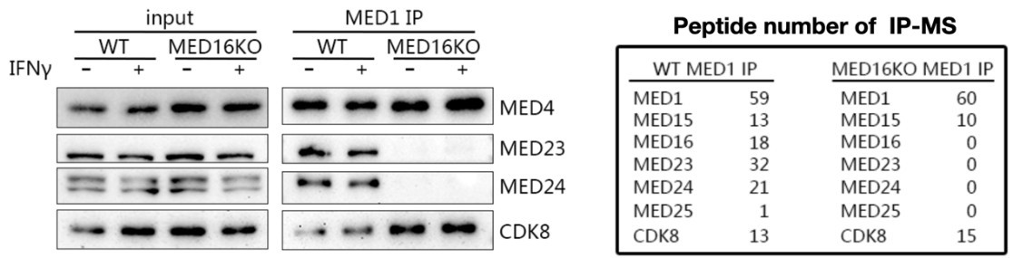
We thank the reviewer for raising this important point. Indeed, our new Co-IP and IP-MS data confirm that MED16 knockout leads to partial dissociation of MED23, MED24, and MED25 from the Mediator complex. We agree that this structural perturbation must be carefully considered when interpreting phenotypes from MED16 depletion experiments, and we acknowledge that long-term knockdown or knockout approaches are inherently susceptible to secondary effects and cellular adaptation.
Author response image 3.
MED16 knockout impacts on the Mediator complex
However, our key conclusions are not based solely on long-term depletion experiments. Several lines of evidence, including experiments performed on short timescales without prolonged MED16 loss, converge on the same conclusion:
First, the MED16 mutant lacking its C-terminal αβ-domain cleanly separates the two functions. This mutant retains the intact N-terminal WDR domain of MED16 and can integrate into the Mediator complex, preserving its structural role and preventing tail module dissociation. Yet it completely fails to exert the competitive inhibition (squelching) effect on the UBP1 reporter that is observed with wild-type MED16 overexpression (Fig 3G), and similarly fails to inhibit HIV-1 reporter activity (Fig 5G). The sole molecular defect of this mutant is its inability to bind UBP1 (Fig 2B). Critically, this overexpression experiment is performed in a wild-type background where the endogenous Mediator complex remains structurally intact. The only variable is whether the overexpressed MED16 can interact with UBP1. This provides strong evidence that the transcriptional effects are mediated specifically through the MED16-UBP1 interaction interface, independent of MED16's structural role in maintaining Mediator integrity.
Second, the transcriptional changes we observe are not random but show specific functional coherence. If the transcriptomic changes in MED16 knockdown cells were merely secondary consequences of Mediator tail module disruption, we would expect broad, non-specific dysregulation. Instead, the UBP1-MED16 co-regulated genes are enriched for specific biological processes—angiogenesis—which largely align with the known biological functions of UBP1 (Fig 4D-E). This functional specificity is difficult to reconcile with the global, non-specific transcriptional disruption that would be expected from the total Mediator dysfunction.
Finally, we wish to address the reviewer's observation regarding Pol II levels in Figure 8B. In preparing the revised manuscript, we carefully re-examined our original Western blot data and found that the reduction of Pol II upon MED16 overexpression was not consistently reproducible across independent experiments. We have therefore replaced new Pol II blot in Figure 8B. We note that this revision does not affect any of the conclusions drawn from Figure 8, which focus on the inhibitory effect of MED16 overexpression on HIV-1 latency reversal in J-Lat cells.
We are grateful for the reviewer’s careful assessment, which encouraged us to refine our data presentation and enhance experimental rigor. The manuscript has been significantly improved as a result.
Reviewer #3 (Public review):
Weaknesses:
(1) The decrease in 1,6-hexanediol-treated cells of MED16 is modest, variable, not quantified, and internally inconsistent. For example, in Figure 1A, 1,6-hexanediol treatment should not have an impact on the level of the protein being directly IP. For MED12 (and CDK8 and MED1 to a lesser extent), 1,6-hexanediol treatment alters the level of the target protein in the IP. Along these lines, Figure 1A shows a no 1,6H-D dependent decrease in MED1 or MED12 levels in the CDK8 IP, whereas Figure 1B does show a decrease. Figure 1A shows no 1,6H-D dependent decrease in CDK8 levels in the MED1 IP, whereas Figure 1B shows a dramatic decrease. MED24 levels in the MED12 IP increase upon 1,6H-D in Figure 1A, but decrease in Figure 1B. Internal inconsistencies of this nature persist in the other Figures.
We thank the reviewer for this careful observation, and we here clarify the rationale behind our experimental design to resolve the apparent discrepancies between Figures 1A and 1B.
We first performed co-immunoprecipitation (co-IP) assays using HEK293T whole-cell lysates with three distinct antibodies targeting CDK8, MED1, and MED12, respectively, to pull down intact Mediator complexes. This setup was designed to assess whether 1,6-hexanediol (1,6-HD) impairs Mediator complex integrity and, if so, whether this perturbation affects individual subunits unequally. Across all three independent IP replicates, we observed a uniform trend: most Mediator subunits displayed only mild signal attenuation, whereas MED16 exhibited a striking, dose-dependent reduction upon escalating 1,6-HD concentrations (Fig 1A). Quantitative densitometry of these immunoblot signals has now been incorporated into the revised Figures 1A and 1B to better visualize this difference. Thus, this initial result in Fig 1A indicated that MED16’s interaction with the core Mediator complex is uniquely vulnerable to 1,6-HD disruption, irrespective of which Mediator subunit was used for immunoprecipitation.
To validate the reproducibility of this phenotype, we replicated the assay in an alternative biochemical system using HeLa nuclear extracts (NEs) (Fig 1B), with 2,5-hexanediol (2,5-HD) included as a critical negative control—this structural isomer cannot disrupt weak hydrophobic protein-protein contacts. Notably, the HeLa NE experiments in Fig 1B were carried out under elevated salt conditions (~300 mM NaCl), in contrast to the 150 mM NaCl buffer used for the HEK293T lysate assays in Figure 1A.
We want to emphasize that despite these substantial technical disparities between the two experimental setups, both datasets converge on the identical core conclusion: MED16’s association with the core Mediator complex is selectively disrupted by 1,6-HD. This consistent trend is further strengthened by quantitative analysis of immunoblot band intensities from biological replicates, which we have added to the revised figures.
Minor variability observed for other Mediator subunits between Fig 1A and 1B arises from intrinsic differences in cell type, extraction protocols, and buffer salt concentrations. For this reason, we did not draw mechanistic interpretations regarding the variable signal changes of other subunits across distinct experimental conditions; instead, our analysis focused on the robust, consistent loss of MED16 following 1,6-HD exposure.
(2) Undermining the value of Figure 1E/F, UBP1 and TFCP2 may also associate with the small amount of MED16 in the 2MDa fractions. This is not tested, and therefore, the conclusion that they just associate with the dissociable form of MED16 is not supported.
We thank the reviewer for this insightful observation. Indeed, UBP1 and TFCP2 can associate with MED16 in the context of the intact Mediator complex. Figure 1B directly supports this: under 2,5-HD conditions, where MED16 remains integrated into the Mediator, both UBP1 and TFCP2 are co-immunoprecipitated with the Mediator complex (via anti-MED1). This association is lost under 1,6-hexanediol conditions, where MED16 dissociates—suggesting that UBP1 and TFCP2 are tethered to the Mediator through MED16.
The gel filtration experiment (Fig 1D) further reveals that a portion of MED16 resolves away from the core Mediator complex under these in vitro conditions, and Figure 1E-F shows that UBP1 and TFCP2 co-immunoprecipitated with MED16 in these lower molecular weight fractions. Taken together, these data indicate that UBP1 and TFCP2 can be found in association with MED16 in two biochemically distinguishable states: one integrated into the Mediator complex, and another that has dissociated from it under our experimental conditions. We did not state that UBP1 and TFCP2 associate exclusively with the dissociated form; rather, the dissociated fraction is where we initially identified this interaction, which then led us to investigate its functional significance. The whole manuscript has been extensively rewritten now, and hopefully the confusion has been clarified.
Our functional data further demonstrate that the transcriptional activities of UBP1 and TFCP2 require the intact Mediator complex. MED16 bridges this interaction through its C-terminal αβ-domain, which binds UBP1/TFCP2, and its N-terminal WDR domain, which anchors MED16 into the Mediator. We have revised the manuscript to clearly distinguish the biochemical observation from the functional model, and we now explicitly state that UBP1 and TFCP2 associate with the Mediator complex through MED16.
(3) Domain mapping studies in Figure 2 are overinterpreted. Since the interactions could be indirect, it is not accurate to conclude "Therefore, the N-terminal WDR domain of MED16 is crucial for its integration into the Mediator complex, while the C-terminal αβ-domain is essential for interacting with UBP1-TFCP2."
We fully acknowledge that co-immunoprecipitation (co-IP) assays alone cannot discriminate direct physical binding from indirect protein associations mediated by intermediate partners. Nevertheless, our conclusion that MED16 directly interacts with UBP1 is supported by two orthogonal, independent experimental datasets.
First, GST pull-down assays using purified recombinant proteins expressed in E. coli confirm direct binding (Figure 2C). This cell-free biochemical system removes all potential cellular bridging factors, unambiguously demonstrating a physical contact between the C-terminal αβ-domain of MED16 and UBP1.
Second, our mutational functional analyses further rule out an indirect interaction model. Deletion of the unique 36-residue USP peptide in UBP1 completely abrogates MED16 binding (Figure 2F, comparison between ΔSAM and DBD truncations). Reciprocally, MED16 mutants lacking the C-terminal αβ-domain lose all capacity to associate with UBP1. Importantly, both truncation mutations yield identical functional defects in UBP1-driven and HIV-1 luciferase reporter assays (Figures 3G and 5G). If this complex assembly relied on an uncharacterized bridging protein, it would be statistically improbable for two distinct, structurally unrelated deletions (USP removal on the UBP1 side and αβ-domain truncation on the MED16 side) to independently disrupt the same indirect linkage and generate identical loss-of-function phenotypes.
Collectively, the congruent results from cell-free in vitro biochemistry and targeted mutational functional assays provide robust evidence for a direct, specific physical interaction between MED16 and UBP1.
(4) A close examination of Figure 2C undermines confidence in the association studies. The bait protein in lanes 5-8 should be equal. Also, there is significant binding of GST to UBP1 and TFCP2, in roughly the same patterns as they bind to GST-MED16 αβ. The absence of input samples makes the results even more difficult to interpret.
We acknowledge the technical limitations the reviewer pointed out. Regarding the concern about unequal protein loading across lanes 5-8, we note that lanes 7 and 8 in fact contain more GST-M16αβ bait protein than lanes 5 and 6, yet TFCP2L1 and YY1 still show no appreciable binding above the GST-alone background. This loading asymmetry therefore strengthens the conclusion that these proteins do not directly interact with MED16. For UBP1 and TFCP2, we have now provided quantification of the Western blot signals, which confirms a clear difference in binding to GST-M16αβ versus GST alone. Furthermore, these GST pull-down experiments were performed under stringent conditions—300 mM NaCl and 0.5% Tween-20 in the wash buffer—and the UBP1 and TFCP2 interactions with GST-M16αβ persisted under these conditions (lane 5-8), whereas binding to GST alone was minimal (lane 1-4). These technical details support the specificity of the observed interactions.
(5) The domain deletion mutants are utilized throughout the manuscript as evidence of the importance of the UBP1-MED16 interaction. However, in Figure 2F lanes 7 and 8, the delta-S mutant binds MED16 as well as full-length UBP1. This undermines much of the subsequent data and conclusions about specificity.
We believe the reviewer may have a misunderstanding about the experimental design. The delta-S (ΔSAM) mutant deletes the SAM motif, which is required for UBP1-TFCP2 heterodimerization, but retains the UBP1-specific sequence (USP) that mediates MED16 binding. The observation that ΔSAM still binds MED16 at levels comparable to full-length UBP1 is therefore not a discrepancy—it is the expected result and confirms that USP is the region responsible for MED16 interaction. The DBD mutant, which further removes USP, completely loses MED16 binding. Together, these deletion constructs delineate two separable interaction surfaces on UBP1: USP for MED16 binding and the SAM motif for TFCP2 heterodimerization (see Figure 2 G). The functional analysis of these domains is reflected in Figure 3B: ΔSAM, which retains MED16 binding but loses TFCP2 dimerization, shows approximately half the transcriptional activity of full-length UBP1, suggesting that both interactions contribute to full UBP1 function. To avoid similar confusion, we have renamed ΔS to ΔSAM,UBP1-SP to USP in the revised manuscript.
(6) Even if the delta-S mutant were defective for MED16 binding, the result in Figure 3B does not "confirm that MED16 is required for the transcriptional activity of UBP1,". Removal of that domain may have other effects.
We wish to first clarify a factual point: the delta-S mutant (ΔSAM) is not defective for MED16 binding. ΔSAM deletes the SAM motif that is required for UBP1-TFCP2 heterodimerization, but retains the UBP1-specific sequence (USP) that mediates MED16 binding. As shown in Figure 2F, ΔSAM binds MED16 at levels comparable to full-length UBP1. The mutant that loses MED16 binding is DBD, which further deletes USP.
The functional result in Figure 3B is therefore consistent with this biochemical mapping: ΔSAM retains MED16 binding but loses TFCP2 dimerization, and accordingly shows approximately half the transcriptional activity of full-length UBP1. This suggests that both the MED16 interaction (via USP) and the TFCP2 interaction (via the SAM motif) contribute to full UBP1 function.
More importantly, the conclusion that MED16 is required for UBP1 transcriptional activity does not rest on domain mutant analysis alone. It is supported by independent loss-of-function approaches: shRNA-mediated MED16 knockdown (Fig 3C) and CRISPR-mediated MED16 knockout across five independent frameshift clones (Fig 3E-F), both of which consistently reduce UBP1 reporter activity. The domain deletion mutants complement these approaches by mapping the specific interaction interfaces.
(7) As Mediator is critical for the activation of many genes, it is not accurate to assume that the impact of its deletion in Figure 3E/F demonstrates a direct requirement in UBP1-driven transcription. This could easily be an indirect effect.
MED16 knockout data must be interpreted cautiously due to potential Mediator destabilization artifacts. However, our conclusion does not rely solely on loss-of-function data; transient competitive overexpression and domain-mapping assays bypass chronic MED16 depletion entirely. The MED16Δαβ mutant retains full Mediator integration but loses UBP1 binding. Unlike wild-type MED16, this mutant fails to change UBP1 or HIV-1 reporter activity in 36–48 h transient assays performed in wild-type cells with intact endogenous Mediator. Dose titration further confirms that only UBP1-interacting MED16 competes for UBP1 binding to repress target promoters (Fig 3G). Since these experiments involve no permanent MED16 ablation, the observed transcriptional effects cannot be attributed to indirect global Mediator dysfunction, verifying specific MED16–UBP1 regulation.
(8) Without documenting the relative protein expression levels in Figure 3G/H, conclusions cannot be drawn about the titration experiments, nor the co-expression experiments. These findings are likely the result of squelching or some form of competition that is not directly related to the UBP1-mediated transcription. A great deal of validation would be required in order to support the model that these effects are a result of MED16 overexpression sequestering UBP1 away from holo-Mediator.
We thank the reviewer for this insightful comment. In fact, the squelching interpretation that the reviewer proposes is fully consistent with our functional model and helps reinforce a key conclusion of our study.
The reviewer correctly points out that overexpressed MED16 may compete with the endogenous Mediator complex for UBP1 binding, sequestering UBP1 away from the holo-Mediator and thereby inhibiting UBP1-driven transcription. This interpretation directly implies that UBP1 requires association with the intact Mediator complex—not merely free MED16—to activate transcription. This is precisely the model we advocate, and the squelching phenomenon observed in Figure 3G thus provides independent functional evidence for it.
The specificity of this effect is confirmed by the MED16 Δαβ mutant: the Δαβ mutant integrates into the Mediator complex normally but cannot bind UBP1, and consequently fails to exert any competitive inhibition on the UBP1 reporter (Fig 3G). This demonstrates that the squelching effect is specifically dependent on the MED16-UBP1 interaction interface. We have now documented the relative protein expression levels for these experiments in the revised manuscript.
(9) The lack of any documentation of expression levels for the various ectopic proteins in the majority of Figures, renders mechanistic claims meaningless (Figures 3, 4, 5, 6, 7, S2, S3). This is particularly relevant since the model presented for many of the results invokes concentration-dependent competition.
In the revised manuscript, we have now included Western blot data documenting the expression levels of ectopic proteins for the key experiments. The expression level data confirm that the proteins were expressed at the expected levels across the different conditions.
Recommendations for the authors:
Reviewing Editor Comments:
The individual reviews are included above. Please also note that additional instructions regarding the publication of the preprint are below.
If you choose to publish your preprint with eLife, these reviews will also be published with the article. As noted in the instructions following the reviews, you do have a chance to include a provisional response to the reviewers' comments in the provisional publication of the preprint. You will also have an opportunity to submit a revised version of the manuscript in the future. Finally, you do have the option of formally withdrawing the article from eLife after the Reviewed Preprint has been published and pursuing publication elsewhere.
Reviewer #1 (Recommendations for the authors):
(1) The writing and presentation are generally good, except that some typos and mistakes in conceptual statements (e.g., lines 407-408 "WDR-deleted MED16, which cannot interact with UBP1") often confuse the readers.
We thank the reviewer for the careful reading. We have tried out best to correct the typos and misstatements, including the error in line 407-408 where "WDR-deleted MED16, which cannot interact with UBP1" should refer to the αβ-domain deletion rather than the WDR deletion. The whole manuscript has been extensively rewritten for better clarity and preciseness.
(2) Inconsistency in the citation format between numbering and author names should be avoided.
The citation format has now been unified throughout the manuscript.
(3) Two important scientific concepts based on the published literature should be mentioned:
(a) "The position-dependent function of transcription factors (TFs) relative to the TSS" (lines 427-428) is a well-known fact in the transcription field that partly explains gene activation versus gene repression; and
(b) the statement "JQ1 that is known to reverse HIV-1 latency by antagonizing BRD4's suppression of the Tat-P-TEFb/SEC (Super Elongation Complex) interaction" (lines 384-386) is only partially correct and is a property unique to the BRD4 long (BRD4-L) isoform.
We thank the reviewer for this suggestion. We have now cited the relevant literature on the position-dependent function of transcription factors relative to the TSS, including the recent study by Duttke et al. (3), which systematically demonstrated that the effect of TF binding on transcription initiation is highly position-dependent. Our findings provide a clear example that UBP1 activates transcription when its binding motif is located upstream of the TSS but represses when the motif overlaps the TSS, which exemplifying this concept.
We have corrected the statement to specify that JQ1 antagonizes the BRD4 long isoform (BRD4-L)-mediated suppression of Tat-P-TEFb/SEC, and have noted that the BRD4 short isoform (BRD4-S) may also contribute to HIV-1 promoter repression and JQ1-mediated latency reversal.
(4) Another relevant concept is that BRD4 short (BRD4-S) isoform-inhibited HIV-1 promoter activity could be potentially alleviated by JQ1, leading to latency reversal. Besides the aforementioned points and those comments already provided in the Public Review, the involvement of TFCP2 and YY1 in MED16-UBP1 function has not been clearly established and will require more careful and detailed investigation.
We thank the reviewer for these suggestions. Regarding BRD4-S, we have added a discussion of the BRD4 short isoform's role in HIV-1 promoter regulation and its potential contribution to JQ1-mediated latency reversal.
Regarding the involvement of TFCP2 and YY1, we agree that their precise roles within the MED16-UBP1 regulatory axis have not been fully dissected in the current study. Our data indicate that TFCP2 can dimerize with UBP1 to enhance transcriptional activation (Fig 3H), and YY1 is present in the MED16-containing fractions (Fig 1E), consistent with its previously reported role in HIV-1 silencing. However, the molecular details of how TFCP2 and YY1 contribute to MED16-UBP1 function—and whether they act in the same or parallel pathways—remain to be determined. We have acknowledged this as a limitation and a direction for future investigation in the revised Discussion.
Reviewer #2 (Recommendations for the authors):
(1) The data from Figure 1F are hard to interpret, the blot quality is poor for Med23, 16
We acknowledge that the blot quality for MED23 and MED16 in Figure 1F was suboptimal in the original submission. We have now provided better Western blot, which confirms the specific co-immunoprecipitation of MED16 and MED23 with TFCP2 in both HeLa nuclear extract and the gel filtration fractions. Importantly, the reciprocal co-IP (anti-MED16 IP blotted for TFCP2 and UBP1, Fig 1E) independently validates this interaction.
(2) Figure 2: No MW markers on gels. Also, these types of experiments are ok for pilot studies, but they aren't rigorous. Only a few proteins were tested, and it's easy to get false positives with how the experiments were completed. Finally, the Mediator proteins were tested as single isolated proteins rather than in their native context. As isolated proteins, they likely have large exposed hydrophobic patches (normally bound to other subunits), and this can generate non-specific "sticky" interactions that can lead to false positive results.
We have now included MW markers on the gel images in the revised Figure 2.
The reviewer raises a valid general concern about domain mapping experiments. However, we wish to emphasize that the readout in Figure 2B and 2F is the binding of exogenously expressed truncated proteins to their endogenous interaction partners—MED1, TFCP2, MED16, and MED23—which are natively folded and assembled in their physiological environment. This is not a situation where two isolated proteins are mixed in a test tube. Furthermore, the deletion series themselves contain important internal controls against non-specific "stickiness": In Figure 2B, progressive deletion of the WDR domains leads to a gradual reduction in MED1 binding, consistent with the predicted structural role of the β-propeller (WDR domain). The truncation at the αβ-domain specifically abolishes TFCP2 binding but does not affect its association with Mediator complex as indicated by MED1. If the truncated proteins were non-specifically sticky due to exposed hydrophobic patches, one would expect uniform binding across all truncations or random binding patterns, which is not observed. And in Figure 2F, deletion of the 36-amino-acid USP abolishes its MED16 binding, while deletion of the SAM motif does not. This sequence-specific differential effect is inconsistent with non-specific hydrophobic interactions.
The domain mapping experiments in Figure 2 were not designed as an unbiased screen but as hypothesis-driven tests guided by the IP-MS results identifying UBP1/TFCP2 as MED16 interactors, sequence and structural analysis predicting the roles of the WDR and αβ-domains. The conclusions from these experiments are corroborated by subsequent functional assays: the USP-deleted mutant and the MED16 Δαβ mutant show corresponding functional defects in both UBP1 reporter and HIV-1 reporter assays (Fig 3G, Fig 5G), providing strong support that the mapped interaction interfaces are functionally relevant.
(3) Figure 3: UBP1 reporter is artificial, 6X UBP1 sites upstream of a TATA box.
We appreciate this comment and wish to clarify a misunderstanding. The UBP1 reporter used in Figure 3 is not an artificial construct assembled from random elements. Its backbone is derived from the mouse α-globin gene promoter, a well-established natural target of UBP1/TFCP2, which contains two endogenous UBP1/TFCP2 binding sites. In order to increase the reporter's sensitivity to UBP1/TFCP2, we expanded the original two sites to six. Critically, this expansion did not alter the native architecture of the promoter—the relative positions of the transcription factor binding sites, the CCAAT box, the TATA box, and the TSS remain unchanged from the original α-globin promoter. This reporter therefore retains the physiological spacing and topological organization of a genuine UBP1 target promoter, and its low basal activity and high inducibility by UBP1 make it a well-suited tool for modeling UBP1 transcriptional activity. We have further clarified this description in the revised manuscript.
(4) Figure 3D: Med16 KD not compelling, perhaps 50% knockdown? Hard to tell because no replicates and no quantitation.
We acknowledge that the shRNA-mediated MED16 knockdown in Figure 3D was partial and that the original presentation lacked replicate quantification. We initially tested three independent shRNAs targeting MED16, of which only one achieved appreciable knockdown. Recognizing this limitation, we turned to a more definitive loss-of-function approach: CRISPR-Cas9-mediated MED16 knockout. Five independent frameshift KO clones were generated in mouse HT cells, each verified by sequencing and Western blotting (Fig 3F), and all five clones consistently showed reduced UBP1 reporter activity compared with wild-type cells (Fig 3E). The consistency between the shRNA knockdown and CRISPR KO reinforce the conclusion that MED16 is required for UBP1 transcriptional activity.
(5) Figure 3E: Med16 KO will be a different cell line because of genetic adaptation. That is, an RNA-seq experiment is expected to result in many gene expression changes compared to the parental line. This could contribute to differences, or differences could result from altered Mediator composition to compensate for Med16 loss, but this wasn't addressed by the authors. A rapid degron method would be much more rigorous.
We thank the reviewer for raising this important concern. We agree that prolonged MED16 loss can elicit cellular adaptation and compensatory changes in Mediator composition, as our own new data on tail module dissociation confirm. This makes it challenging to disentangle direct and indirect effects using constitutive KO models alone.
However, two independent lines of evidence support a direct role for MED16 in UBP1-mediated transcription that cannot be explained by adaptive responses:
First, acute shRNA-mediated knockdown produces the same phenotypic trend. shRNA operates on a shorter timescale (days) without prolonged selection for compensatory mutations, yet it also reduces UBP1 reporter activity (Fig 3C). The convergence of acute and chronic loss-of-function approaches suggests the phenotype is driven by MED16 loss itself rather than by the secondary adaptations that accumulate over time in KO cells.
Second, and most critically, the MED16Δαβ mutant experiment completely bypasses the issues of genetic adaptation and Mediator structural disruption (Fig 3G). This experiment is performed in wild-type cells with an intact endogenous Mediator complex, without any MED16 depletion and without any long-term selection. The MED16Δαβ mutant retains full ability to integrate into the Mediator complex (its WDR domain is intact) but cannot bind UBP1. Overexpression of wild-type MED16 competitively inhibits the UBP1 reporter due to the squelching effect, whereas overexpression of MED16Δαβ at comparable levels does not (Fig 3G). The sole molecular difference is that MED16Δαβ doesn`t contain the UBP1-binding interface. Because this experiment involves no MED16 loss and no adaptive period, the result directly demonstrates that MED16-UBP1 interaction is required for UBP1 transcriptional activity, entirely independent of concerns about clonal adaptation.
We appreciate the reviewer's suggestion of a rapid auxin-inducible degron approach. While acute depletion strategies offer clear advantages in minimizing cellular adaptation, they are not without limitations. First, degron-based depletion rarely achieves 100% elimination of the target protein, and residual protein may confound interpretation. Second, and particularly relevant to our system, degron-mediated degradation may preferentially target free, unassembled subunits while sparing proteins already stably integrated into large macromolecular complexes such as the Mediator—potentially creating a lag or even complete resistance to degradation for the pool of MED16 that is assembled into the complex. Given these considerations, we believe our convergent strategy—combining shRNA knockdown, CRISPR-mediated knockout in multiple independent clones, and the Δαβ mutant that bypasses structural perturbation—provides a robust and complementary set of evidence that does not rely on any single depletion method.
(6) The title for Figure 3 is "Med16 is required for UBP1 activation of gene transcription", but that's not conclusively shown by these experiments.
We agree that the original title of Figure 3 may overstate. We have revised the title to " MED16 acts through the Mediator complex to support UBP1-mediated transcriptional activation ", which may better reflect the data. As discussed in our responses above, this conclusion is supported by converging evidence from shRNA knockdown (Fig 3C), CRISPR-mediated KO across multiple clones (Fig 3E-F), and the specific loss of function observed with the MED16Δαβ mutant (Fig 3G).
(7) Figure 5: Med16 protein levels were never measured, nor were other transfected proteins. Reporter assay data are correlative only; no evidence for direct effects, and only 2 other MED subunits are probed. Again, this is a starting point, but nothing to use for concrete conclusions about Med16.
We have now provided Western blot data documenting MED16 and other transfected protein levels for the experiments in Figure 5, confirming that the proteins were expressed at the expected levels. We agree that plasmid-based reporter assays alone cannot establish direct transcriptional mechanisms. The HIV-1 reporter experiments in Figure 5 serve as an initial functional readout, and their conclusions are corroborated by more direct approaches: (i) the position-dependent specificity demonstrated by chimeric reporter assays (Fig 6A-C); (ii) the independence of the effect from TAR-mediated elongation and HDAC activity (Fig S2), ruling out alternative mechanisms; and (iii) the immobilized template transcription assays, which directly measure nascent RNA production and PIC assembly on the HIV-1 promoter (Fig 7). The reporter data should be viewed as one component of a larger body of convergent evidence rather than as standalone proof.
MED23 and MED24 were chosen as the most relevant controls because they, along with MED16, form the core of the tail module submodule that creates the transcription factor interaction platform. They are structurally adjacent to MED16 within the same submodule. The fact that neither MED23 nor MED24 overexpression affects HIV-1 reporter activity (Fig 5C), whereas MED16 overexpression does so in a UBP1-binding-dependent manner (Fig 5C, 5E), demonstrates that the inhibitory effect is specific to MED16 and not a generic consequence of overexpressing a Mediator tail subunit.
(8) Figure 6: similar concerns to Figure 5. The promoters are artificial, derived from a chicken beta-actin promoter with artificially inserted UBP1 binding sequences.
We acknowledge that the chimeric promoters in Figure 6 are indeed deliberately designed. To address this specific question: Does the binding position of the UBP1 relative to the TSS dictate whether UBP1-MED16 activates or represses transcription?
To isolate position as the sole variable, we needed a backbone promoter that (i) is stably and robustly active in the cell lines used, (ii) does not itself contain endogenous UBP1 binding sites that would confound the interpretation, and (iii) allows precise placement of the UBP1 binding motif either upstream of or overlapping the TSS. The chicken β-actin promoter meets all three criteria and is a widely used tool in molecular biology for such controlled promoter engineering experiments. Notably, its high GC content around the promoter region is also a feature shared with the HIV-1 core promoter, making it a particularly relevant backbone for the HIV-1 TSS chimeric constructs.
The key controls for this experiment are internal: in the same backbone, placing the UBP1 binding site at the TSS results in MED16-UBP1-dependent repression, whereas placing it upstream does not (Fig 6A vs 6B). Furthermore, this position-dependent effect requires the MED16-UBP1 interaction interface, as the MED16 Δαβ mutant and the UBP1 DBD mutant both fail to produce the inhibitory effect (Fig 6A). The differential outcome between the two constructs—which differ only in the placement of the UBP1 binding site—cannot be attributed to the artificial nature of the backbone, because the backbone is identical in both cases.
(9) Figure 6D: Not rigorous, no statistical analysis, cannot determine whether the modest changes are biologically relevant/meaningful. Also, data were obtained through indirect measurements and long timeframes, so any direct effects from possible UBP1 binding are probably lost.
The differentially expressed genes in Figure 6D were selected using DESeq2 with a significance threshold of p < 0.05. The top 50 upregulated and top 50 downregulated transcripts were then used for UBP1 motif enrichment analysis around their promoters. We have clarified these selection criteria in the revised figure legend.
Regarding the concern that indirect measurements over long timeframes, we now have additional evidence that UBP1 binding effects are preserved in our RNA-seq data. Motif analysis of UBP1-upregulated genes using HOMER identified the CP2 family binding motif (CNRG-N6-CNRG) as the most significantly enriched motif. This indicates that UBP1-binding-dependent transcriptional effects are not lost in our dataset.
Author response image 4.
Motif enrichment result of the UBP1 up-regulated genes
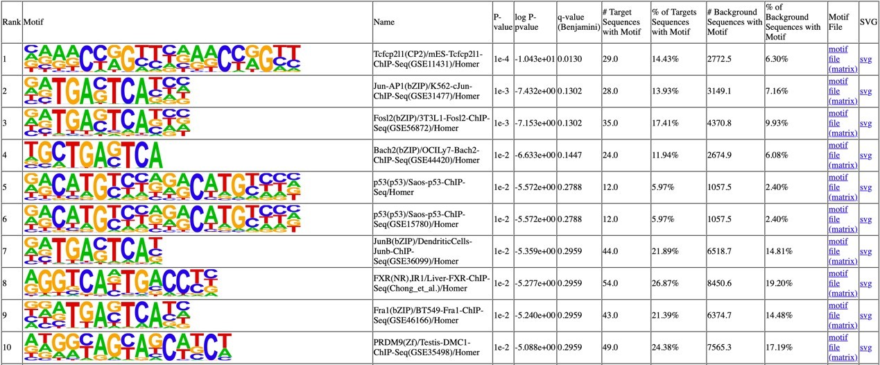
Furthermore, when these UBP1-upregulated and UBP1-downregulated genes are analyzed separately for motif position relative to the TSS, a clear pattern emerges: UBP1-upregulated genes show motif enrichment flanking the TSS, whereas UBP1-downregulated genes show enrichment overlapping the TSS (Fig 6D). If the transcriptional changes were non-specific secondary consequences of prolonged UBP1 overexpression, one would not expect this position-specific distribution. Finally, this positional specificity is corroborated by the chimeric reporter experiments (Fig 6A-C), where moving the UBP1 binding site across the TSS in an otherwise identical promoter directly switches the regulatory outcome. The concordance among the de novo motif discovery, the genome-wide positional analysis, and the functional reporter assays supports the biological relevance of the motif enrichment pattern.
(10) The title for Figure 6 is "UBP1 binding site determines the UBP1-Med16 inhibition in HIV-1 transcription" but the data do not convincingly show this.
We appreciate this suggestion. We have revised the title to "The position of the UBP1 binding site relative to the TSS dictates whether MED16-UBP1 activates or represses transcription.". For clarification, Figure 6 not only demonstrates that the UBP1 binding position dictates MED16-UBP1-mediated inhibition in the HIV-1 context, but also revealed that this positional effect could be generalized to endogenous genes: UBP1 motifs flanking the TSS are associated with activation, while motifs overlapping with the TSS are associated with repression.
(11) Figure 7: The data and the experiment do not support the claim in the title that "MED16-UBP1 complex prohibited PIC formation of HIV-1 transcription". I cannot convince myself that there are meaningful differences between the results shown in 7B, but it's hard to judge because there are no replicates and no quantitation.
We have revised the title from "MED16-UBP1 complex prohibited PIC formation of HIV-1 transcription" to "MED16 interferes with HIV-1 PIC assembly at the TSS via its interaction with UBP1." We have also provided quantification of the Western blot signals from replicate experiments in Figure 7B, which confirms statistically significant differences in CDK8 binding, Pol II pSer5 signals, and TFIIB retention between conditions. We note that the key observations in Figure 7B were consistently observed across multiple pilot experiments prior to the final dataset presented in the manuscript. Thus, the reported differences are reproducible and not based on a single experiment.
(12) The immobilized template experiments had only -78 upstream and +60 downstream of the TSS. By my understanding, much more upstream DNA is needed to allow a PIC to assemble, because the 5'-end bead attachment will block. I think this was outlined in an article by the Carey lab, but I'm not sure. A point here is that it would be reassuring for the authors to show that PIC assembly was occurring and that transcription was responsive to a transcription factor, similar to what the Carey lab has done with immobilized templates. See, for example, Figure 1 in Lin & Carey Curr Protocol Mol Biol 2012 Ch12 unit 12.14.
We thank the reviewer for raising this technical consideration. We respectfully offer a different perspective based on current structural data on the human PIC.
A recent cryo-EM study of the human Mediator-bound preinitiation complex (4) demonstrated that the PIC spans approximately 50 bp upstream and downstream of the TSS. The DNA templates used for structural determination in that study were generally within 105 bp upstream and 63 bp downstream of the TSS. Our HIV-1 template (-78 to +60, i.e., 78 bp upstream and 60 bp downstream of the TSS) falls within this well-characterized range and provides sufficient space for PIC assembly.
Regarding the choice of the -78 boundary: the HIV-1 core promoter is defined as -78 to +60, and the region from -78 to -105 encompasses the HIV-1 enhancer, which contains additional regulatory elements including NF-κB and NFAT binding sites. We deliberately excluded these enhancer elements to avoid confounding regulatory inputs and to focus specifically on the UBP1 binding site at the TSS, which is the central focus of our model. The robust luciferase mRNA production observed in wild-type nuclear extract (Fig 7C) provides direct functional evidence that PIC assembly and transcription initiation proceed efficiently on this template under our conditions.
Author response image 5.
Promoter templates used for human PIC assembly (Chen et al., Science 372, eaba8490, 2021): templates within −105 to +65 bp of the TSS support PIC formation
(13) Related to the above concern, the qPCR analysis from the extract experiments may be misleading because of contaminating nucleic acids in the extract. More control experiments are needed, such as no NTP controls.
We thought this concern has been addressed by our experimental design.
First, the PIC step in our experimental design (Fig 7B, "PIC") is the no-NTP control: nuclear extract is incubated with the immobilized template without NTPs. After washing, the proteins bound at this step are analyzed by Western blot, and importantly, there is no transcription occurred at this step. Transcription is then initiated by adding NTPs (Fig 7B, "Trx."), and the supernatant from this step is collected for qPCR analysis of transcription.
Second, it is unlikely that the contaminating nucleic acids from the nuclear extract could confound our qPCR results, because the qPCR analysis specifically detects Luciferase mRNA transcribed from the immobilized HIV-1-Luciferase template, which is absent from eukaryotic genomes; and the endogenous genomic DNA or RNA cannot generate a false-positive signal.
Lastly, after transcription, the immobilized templates are retained on the streptavidin beads and removed, while only the supernatant containing newly synthesized RNA is collected for qPCR. Any residual template DNA or extract-derived nucleic acids bound to the beads are therefore excluded from the qPCR sample.
(14) Some references are made with the format of (author name, year), but others use numbers.
The citation format has been unified throughout the manuscript.
(15) On line 111-115, it is stated that 2,5 HD somehow "does not interfere with weak hydrophobic interaction due to different positions of hydroxyls" with no reference. Both 1,6 HD and 2,5 HD are hydrophobic molecules, and they will disrupt protein structure. The authors make a point about the differential results with 1,6 HD vs. 2,5 HD, but it's not compelling and not justified.
We have now provided the reference to support this statement. The specificity of the 1,6-HD effect is further clarified as follows.
1,6-HD and 2,5-HD are structural isomers sharing the same chemical formula and molecular weight. The key difference lies in the positions of their hydroxyl groups (See Author response image 6). The position of the two hydroxyl groups changes both the molecule’s shape and how it interacts with water and hydrophobic surfaces.
In 1,6-HD, the two -OH groups sit at the ends of the chain, giving a more symmetric, flexible molecule, while in 2,5-hexanediol the -OH groups are closer to the middle, which changes polarity distribution and local geometry (5, 6). The distinctive structures matter because hydroxyl groups strongly hydrogen-bond with water. When they are terminal and well separated in 1,6-HD, the molecule can present a more balanced hydrophilic “cap” at both ends while keeping a relatively long hydrophobic center, which helps 1,6-HD partition into and disturb weakly interacting hydrophobic regions in condensates or hydrophobic interfaces (Author response image 6). By contrast, moving the hydroxyls inward in 2,5-HD does not help with disrupting the hydrophobic interfaces (Author response image 6). That is why 2,5-HD is often used as a comparison compound: it has the similar chemical formula, but the different hydroxyl pattern usually makes it a weaker or at least differently acting perturbant.
Author response image 6.
Differential disruption of hydrophobic interfaces by 1,6-HD vs 2,5-HD
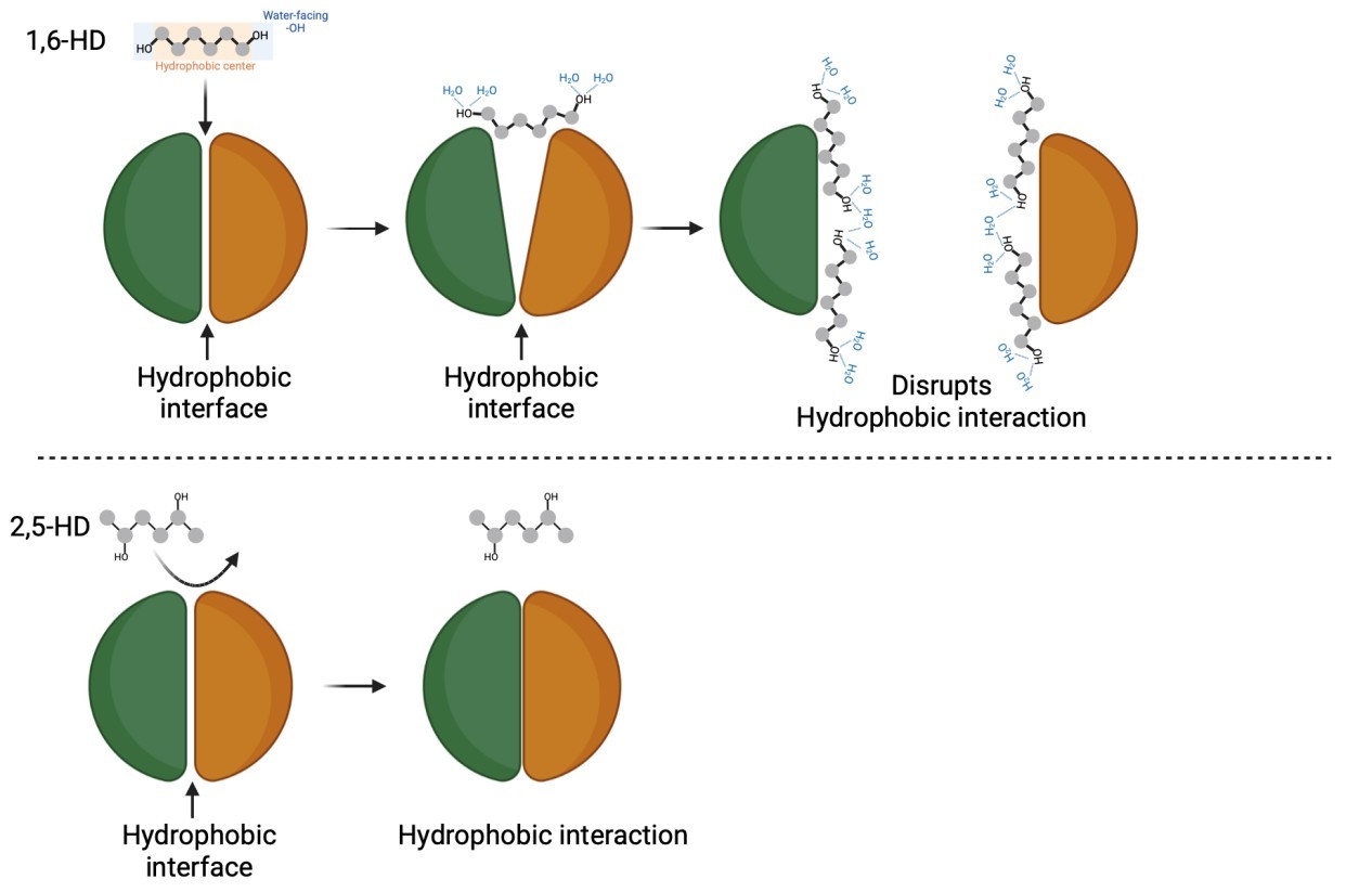
(16) On lines 117-120, the authors claim that Med16 is "integrated into the Mediator complex through weak hydrophobic interaction but not ionic interaction" based upon a few IP results. They then state (lines 130-131) that "the interaction between Mediator and Med16 is an unstable hydrophobic interaction." These statements make no sense based on any data shown, and they ignore the fact that Med16 purifies with Mediator from many different purification protocols. It's safe to say that its interaction with Mediator results from strong, extensive hydrophobic interfaces, which is further supported by actual structural data. The authors should check their reasoning here because they are over-interpreting their results, and this will be misleading to readers.
We thank the reviewer for this important suggestion. The reviewer is right that our original phrasing—particularly "unstable hydrophobic interaction"—was imprecise and inconsistent with the established structural data. As the reviewer notes, MED16 co-purifies with the Mediator complex through multiple purification protocols, and recent cryo-EM structures (4) reveal that MED16 engages in extensive hydrophobic interfaces with neighboring tail module subunits, particularly MED24 and MED25. We fully agree that this is a strong, hydrophobic interaction in the physiological context.
Our 1,6-hexanediol experiments demonstrated that the MED16-Mediator interface does have a hydrophobic character, which renders the sensitivity to 1,6-HD disruption. This does not mean the interaction is weak. We have now carefully described the relevant statements in the revised manuscript.
Reviewer #3 (Recommendations for the authors):
(1) It is not accurate to claim that "198 (genes) were repressed by shMED16 knockdown (Fig 4B and 4C)". These genes were simply not activated by UBP1 in the knockdown cells.
We thank the reviewer for this careful distinction. We have revised the text to state that the UBP1-activated genes were “profoundly downregulated upon MED16 depletion” rather than “repressed by shMED16 knockdown”. The biological point remains unchanged: MED16 is required for UBP1 to activate this set of target genes.
(2) Like the majority of experiments presented, the RNA-seq presented in Figure 4 may reflect indirect effects of MED16 loss. As such, the results do not support the claim that "This result demonstrated that UBP1 and MED16 collaborate to regulate transcription".
We acknowledge that RNA-seq data from MED16 knockdown cells alone cannot distinguish direct from indirect effects. However, we did not rely on the RNA-seq data alone to support a collaborative role for UBP1 and MED16.
The key evidence comes from experiments comparing wild-type MED16 with the MED16Δαβ mutant. MED16Δαβ integrates into the Mediator complex normally but cannot bind UBP1 (Fig 2B). When overexpressed in wild-type cells, wild-type MED16 inhibits the UBP1-driven reporter activity through a squelching effect. In contrast, MED16Δαβ has no effect on the UBP1 reporter assay (Fig 3G), and the same is true for the HIV-1 reporter assays (Fig 5G). Thus, MED16 must both integrate into the Mediator complex and directly bind UBP1 to support UBP1-mediated transcription.
In addition, the genes identified as UBP1-MED16 co-regulated by RNA-seq are enriched for specific biological processes—angiogenesis (Fig 4D-E)—that align with the known physiological functions of UBP1. Such functional coherence would be unexpected if the transcriptional changes were merely non-specific secondary consequences of Mediator disruption.
Thus, while we agree that RNA-seq alone has limitations, the convergent evidence from the MED16Δαβ mutant and the functional specificity of the co-regulated gene set supports a collaborative role for UBP1 and MED16 in transcriptional regulation.
(3) The data in Figure 4 are not presented with controls documenting the relative expression levels of MED16 and UBP1. For example, it would be important to understand the level of UBP1 overexpression achieved, relative to endogenous UBP1 levels in HeLa cells.
We agree with the reviewer that documenting expression levels is important. Author response image 7 are the CPM values for UBP1 from our RNA-seq data, confirming robust overexpression in the UBP1-overexpression condition compared with the control. The MED16 protein levels in the knockdown cells can be found in Fig 3D, as the same MED16-knockdown HeLa cell line was used for the RNA-seq experiment.
Author response image 7.
UBP1 expression level derived from RNA-seq
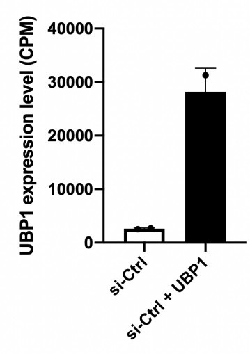
Regarding the physiological relevance of UBP1 overexpression, several observations support that the transcriptional effects we observe are specific rather than artifacts of overexpression: (i) the UBP1-activated genes are enriched for pathways known to be regulated by UBP1—angiogenesis (Fig 4D-E)—rather than showing random, non-specific activation; (ii) the activation of these genes is attenuated upon MED16 knockdown, demonstrating dependence on the MED16-UBP1 interaction; and (iii) in reporter assays, the MED16 mutant lacking its C-terminal αβ-domain fails to competitively inhibit UBP1 driven reporter transcription even when UBP1 is similarly overexpressed (Fig 3G), indicating that the competitive inhibition of the UBP1 reporter depends specifically on the MED16-UBP1 interaction interface, rather than on the amount of UBP1 protein overexpressed.
(4) The enhancer-promoter architecture of the luciferase construct used in Figure 3 is not sufficiently described. Are the elements far enough apart that Mediator-dependent looping is required for transcription, for example?
We appreciate the reviewer's question. The UBP1 reporter construct used in Figure 3 is derived from the mouse α-globin gene promoter, a well-established natural target of UBP1/TFCP2(7, 8). The native α-globin promoter contains two endogenous UBP1/TFCP2 binding sites; to increase the reporter's sensitivity to UBP1, we expanded these to six tandem copies. Critically, this expansion preserved the native architecture of the promoter—the relative positions of the UBP1 binding sites, the CCAAT box, the TATA box, and the TSS were not altered. The entire reporter construct spans only 198 bp in total length. At this scale, all regulatory elements reside within a compact proximal promoter region, with no element separated by a distance that would require DNA looping for functional communication. The reporter therefore measures UBP1-dependent transcriptional activation from the proximal promoter, consistent with the canonical role of Mediator in facilitating PIC assembly rather than mediating long-range enhancer-promoter interactions. We have added a detailed description of the construct architecture to the Methods section.
(5) The identification of matches to the UBP1 consensus sequence in a promoter does not prove that this is how UBP1 overexpression is impacting their transcription. "highly matched UBP1 motifs were identified in their promoters (Fig. 4F), leading to their activation when UBP1 was overexpressed."
We agree with the reviewer that the presence of a UBP1 consensus motif in a promoter does not by itself prove direct regulation. We have revised the original statement: the activation of these genes by UBP1 overexpression is now described as "associated with" the presence of UBP1 motifs in their promoters.
We acknowledge that the ideal experiment to demonstrate direct UBP1 occupancy would be ChIP-seq. However, ChIP-grade antibodies against UBP1 are not commercially available, and no UBP1 ChIP-seq datasets exist in public databases. We attempted to carry out the ChIP-seq ourselves but were unable to obtain sufficiently good-quality data.
In the absence of ChIP data, three lines of evidence indicate that the identified UBP1 motifs are functionally relevant. First, motif analysis of UBP1-upregulated genes using HOMER identified the CP2 family binding motif (CNRG-N6-CNRG) as the most significantly enriched motif. This demonstrates that UBP1-binding-dependent transcriptional could be preserved in our RNA-seq data. Second, when we examine the positional distribution of these motifs, a clear pattern emerges: UBP1 motifs flank the TSS in activated genes, but overlap the TSS in repressed genes (Fig 6D). Indirect effects would not produce this position-specific pattern. Third, our chimeric reporter experiments (Fig 6A-C) directly test this position effect: moving the UBP1 binding site upstream of the TSS leads to activation, while moving it to overlap the TSS leads to repression. Together, these three independent approaches support the conclusion that UBP1 regulates these genes through the motifs we identified. See Author response image 4.
(6) Lines 266 and 277 are redundant.
We have carefully reviewed lines 266 and 277. Line 266 is the topic sentence (caption) of the section ("MED16 collaborates with UBP1 to inhibit HIV-1 transcription"), which introduces the overall conclusion, while line 277 describes the specific experimental result ("MED16 inhibited HIV-luciferase activity in a dose-dependent manner"). These serve distinct rhetorical functions. We have ensured that no other redundant phrasing exists elsewhere in the section.
(7) The mere correlation of binding site position with activation status does not prove "that UBP1 may act as either a transcriptional activator or repressor, depending on distance of its binding site to TSS."
We appreciate the reviewer's concern regarding the distinction between correlation and causation. The genome-wide analysis (Fig 6D) reveals an association between UBP1 motif position and transcriptional outcome, which prompts us to postulate that the UBP1 binding position could dictate the transcriptional activity level. The causal evidence comes from the chimeric reporter experiments (Fig 6A-C), in which we directly manipulated the position of the UBP1 binding site relative to the TSS in an otherwise identical promoter background. Placing the UBP1 binding site at the TSS resulted in MED16-UBP1-dependent repression, whereas placing it upstream of the TSS did not. This controlled experiment demonstrates that changing only the position of the UBP1 binding site is sufficient to switch the regulatory outcome, thereby establishing a causal relationship between binding site position and transcriptional effect. We have revised the text to clearly distinguish the correlational genome-wide analysis from the causal reporter experiments.
(8) In Figure 7, MED16 overexpression results in less CDK8 binding. This undermines the claim that MED16 overexpression lowers transcription by binding to UBP1, and suggests an indirect effect.
We appreciate the reviewer's careful observation. The reduction in CDK8 binding upon MED16 overexpression is consistent with our model: MED16, through UBP1 occupying the TSS, interferes with PIC assembly, of which CDK8 is a component. Importantly, the input levels of CDK8 and other Mediator subunits remained unchanged across the three conditions (Fig 7B), indicating that the reduced CDK8 signal in the PIC is not due to altered protein expression.
In support of the specificity of the transcriptional effect, MED16Δαβ—which integrates into the Mediator complex but cannot bind UBP1 (Fig 2B)—fails to competitively inhibit either the UBP1 reporter (Fig 3G) or the HIV-1 reporter (Fig 5G), confirming that the transcriptional outcome depends on the MED16-UBP1 interaction.
(9) Figure 8F is not referenced in the text or legend.
Figure 8F has now been referenced in the Results and Discussion section.
Reference:
(1) Wang G, Cantin GT, Stevens JL, Berk AJ. Characterization of mediator complexes from HeLa cell nuclear extract. Mol Cell Biol. 2001;21(14):4604-13.
(2) Yamazaki T, Souquere S, Chujo T, Kobelke S, Chong YS, Fox AH, et al. Functional Domains of NEAT1 Architectural lncRNA Induce Paraspeckle Assembly through Phase Separation. Molecular Cell. 2018;70(6):1038-+.
(3) Duttke SH, Guzman C, Chang M, Delos Santos NP, McDonald BR, Xie J, et al. Position-dependent function of human sequence-specific transcription factors. Nature. 2024;631(8022):891-8.
(4) Chen X, Yin X, Li J, Wu Z, Qi Y, Wang X, et al. Structures of the human Mediator and Mediator-bound preinitiation complex. Science. 2021;372(6546).
(5) Zheng T, Wake N, Weng SL, Perdikari TM, Murthy AC, Mittal J, et al. Molecular insights into the effect of 1,6-hexanediol on FUS phase separation. EMBO J. 2025;44(10):2725-40.
(6) Romero CM, Páez MS, Miranda JA, Hernández DJ, Oviedo LE. Effect of temperature on the surface tension of diluted aqueous solutions of 1,2-hexanediol, 1,5-hexanediol, 1,6-hexanediol and 2,5-hexanediol. Fluid Phase Equilibr. 2007;258(1):67-72.
(7) Lim LC, Swendeman SL, Sheffery M. Molecular cloning of the alpha-globin transcription factor CP2. Mol Cell Biol. 1992;12(2):828-35.
(8) Chae JH, Kim CG. CP2 binding to the promoter is essential for the enhanced transcription of globin genes in erythroid cells. Mol Cells. 2003;15(1):40-7.



