
Peer review process
Revised: This Reviewed Preprint has been revised by the authors in response to the previous round of peer review; the eLife assessment and the public reviews have been updated where necessary by the editors and peer reviewers.
Read more about eLife’s peer review process.Editors
- Reviewing EditorLaura ColginUniversity of Texas at Austin, Austin, United States of America
- Senior EditorLaura ColginUniversity of Texas at Austin, Austin, United States of America
Reviewer #1 (Public review):
Summary:
This manuscript by Joshi and colleagues demonstrates that the precise theta-phase timing of spikes is causal for CA1 hippocampal theta sequences during locomotion on a linear track and is necessary for learning the cognitively demanding outbound component of a hippocampus-dependent alternation task (W-maze), independently of replay during immobility. To reach these conclusions, the authors developed a theta-phase-specific, closed-loop manipulation that used optogenetic activation of medial septal parvalbumin (PV) interneurons at the ascending phase of theta during locomotion. This protocol preserved immobility periods, allowing a clean and elegant dissociation from SWR-associated replay.
The manuscript is well written and was a pleasure to read. The work described if of high quality and introduces several notable advances to the field:
a) It extends prior studies that manipulated theta oscillations by examining precise temporal structure (specifically theta sequences) rather than only LFP features.
b) The closed-loop manipulation enabled dissociation between deficits in theta sequences during a behavioural task and SWR-associated replay activity.
c) As controls, the authors included rats with suboptimal viral transduction or optic-fibre placement, and, within subjects, both stimulation-on (stim-on) and stimulation-off (stim-off) trials. Notably, sequence disruption persisted into stim-off periods within the same session.
Overall, this is a strong manuscript that will provide valuable insights to the field.
After revision, the manuscript has been substantially strengthened. The authors did incorporate the vast majority of the reviewer's comments and have expanded the discussion of prior medial septal manipulations, clarified the rationale for their theta-sequence analyses, added analyses of SWR and replay in the rest/sleep box as well as provided additional methodological and histological validation.
The new rest-box analysis is a great addition and directly addresses my request to distinguish aSWRs from events during longer off-track rest periods (rSWR). The data support the narrower conclusion that no large group difference was detected in rest-box ripple rate or duration.
The new observation (in response to reviewer #2, point3.2) that on the W-track theta power does not fully recover during stimulation-off periods does change the interpretation of the results. It means that these epochs are then not a physiologically recovered control condition. Therefore, the persistent disruption of theta sequences during the middle block cannot, alone, demonstrate that disrupting sequences during the earliest experience produced a lasting plasticity-related effect. It could also reflect a lingering network effect of the stimulation that persists after laser delivery has stopped. In my opinion the discussion should present at least these two alternatives: the disruption of early experience-dependent plasticity, as well as the incomplete physiological recovery from the preceding stimulation. The linear-track recovery data is helpful, but it does not guarantee the same mechanisms/effects will be present on the novel Wmaze (versus the familiar linear track).
Reviewer #2 (Public review):
Summary:
The authors of this study developed a closed-loop optogenetic stimulation system with high temporal precision in rats to examine the effect of medial septum (MS) stimulation on the disruption of hippocampal activity at both behavioral and compressed time scales. They found that this manipulation preserved hippocampus single-cell-level spatial coding but affected theta sequences and performance during a spatial alternation task. The performance deficits were observed during the more cognitively demanding component of the task and even persisted after the stimulation was turned off. However, the effects of this disruption were confined to locomotor periods and did not impact waking rest replay, even during the early phase of stimulation-on. Their conclusion is consistent with previous findings from the Pastalkova lab, where MS disruption (using different methods) affected theta sequences and task performance but spared replay (Wang et al., 2015; Wang et al., 2016). However, it differs from a recent study in which optogenetic disruption of EC inputs during running affected both theta sequences and replay (Liu et al., 2023).
Strengths:
The experiments were well designed and controlled, and the results were generally well presented.
Comments on revised version.
The authors of this study addressed all my concerns, some of them successfully. The stimulation disrupted theta oscillations, making quantification of theta sequences problematic. Within the constraints of their experimental design, the authors tried their best to address my concerns. Therefore, I am satisfied with the current version and express no further comments.
Author response:
The following is the authors’ response to the original reviews.
eLife Assessment:
This important study employs a closed-loop, theta-phase-specific optogenetic manipulation of medial septal parvalbumin-expressing neurons in rats and reports that disrupting theta-timescale coordination impairs performance of challenging aspects of spatial behaviors, while sparing hippocampal replay and spatial coding in hippocampal place cells. The findings are expected to advance theoretical understanding of learning and memory operations and to provide practical implications for the application of similar optogenetic approaches. The experiments were viewed as technically rigorous, but the strength of evidence provided in the current version of the manuscript was viewed as incomplete, mostly due to limited analyses and the descriptions of some of the experimental protocols.
We thank all reviewers for their overall assessment, thoughtful comments, and suggestions. We have now addressed each of the reviewers’ comments in detail and updated the manuscript on bioRxiv (URL: https://www.biorxiv.org/content/10.1101/2025.09.15.675587v2). In addition, we have shared the raw data, intermediate analysis files, and the complete repository to facilitate replication of the analysis and figures.
Code repo: github.com/LorenFrankLab/ms_stim_analysis
Data repo: dandiarchive.org/dandiset/001634
Docker containers (see GitHub repo for use instructions):
- Database: https://hub.docker.com/r/samuelbray32/spyglass-db-ms_stim_analysis
- Python notebooks: https://hub.docker.com/r/samuelbray32/spyglass-hub-ms_stim_analysis
(1) Novelty and contrast with earlier manipulations:
We now explicitly contextualize our results with prior pharmacological (Wang et al., 2016; Wang et al., 2015; Koenig et al., 2011; Brandon et al., 2014), systemic (Robbe & Buzsaki 2009; Petersen and Buzsáki 2020), and behavioral (Drieu et al., 2018) manipulations that also assessed some of the physiological features we evaluated. This contrast helps us highlight both the insights and the discrepancies observed in the prior approaches. We also more clearly explain the novelty and importance of our specific approach for temporally and physiologically precise manipulation. Specifically, our approach (closed-loop theta-phase stimulation during locomotion) provides a level of physiological specificity that enables dissociation of theta-state dynamics from other hippocampal processes. This, in turn, allows us to address a question that has remained unresolved across prior studies: Are hippocampal spatial sequences during locomotion (i.e., theta sequences) necessary for learning a novel hippocampal-dependent task?
(2) Additional analysis on SWRs during rest:
Since submitting the manuscript, we have conducted additional analysis on the rate and length of SWRs in the rest box (rSWRs) and found that the rate and length are also indistinguishable between targeted and control animals (effect of manipulation between control and targeted animals; rSWR rate: p=0.45; rSWR length: p=0.94, mixed-effects model). We also find evidence for sequential neural representations (“significant replay”) in the rest box when the encoding was performed in the behavioral arena. Example trajectories and full analysis per animal are shown in the new supplementary figure (Figure S6). These results are consistent with our observations on aSWR rate, length, and content in the behavioral arena. Additionally, based on the reviewer’s recommendation, we have evaluated the fraction of ripples with continuous trajectories during the rest box before the W-Track experience and in the subsequent sleep epochs after the first exposure. We find that with experience on the track, the proportion of continuous replays increases on average in both control and transfected animals, and both groups of animals show an overlapping range of continuous trajectory lengths.
(3) Theta sequence measurement in the absence of theta:
We now explicitly explain why our manipulation makes it more appropriate to measure sequential hippocampal representations during locomotion (i.e., theta sequences) without using theta oscillation or an epoch-averaged, relatively large sliding window as a reference. The key insight here is that our manipulation suppresses theta and thus makes it difficult or impossible to accurately identify theta phase. We explain that while theta-phase-based approaches were used in prior work; these prior analyses may have confounded the absence of hippocampal theta sequences during locomotion by the inability to detect theta oscillatory phase reliably. We show that our method of using clusterless Bayesian decoding, in which we estimate the decoded position at every 2ms timestep, is indeed able to capture endogenous hippocampal sequences even without imposing any requirements of aligning to theta oscillations, thus providing an unbiased estimate of the rhythmicity of hippocampal spatial representations.
(4) Additional analysis on place cell stability and tuning:
We thank the reviewers for this question. For the KL divergence analysis, we have imposed a spike-count criterion (100 spikes for each interval type —stimulation-off, stimulation-on, and the stimulus sub-interval) and a coverage criterion (50% HPD of the units’ spatial firing distribution was contained within 40cm on the linear track and 100cm on the w-track). These criteria were chosen to ensure that spatial tuning curves were sufficiently well sampled and localized to allow reliable estimation of KL divergence, which is particularly sensitive to noise arising from low spike counts or diffuse firing. Based on the reviewer’s suggestion, we have relaxed the unit inclusion criteria for KL divergence by relaxing the criteria for the number of spikes (50 spikes) to include more weakly tuned place cells and replicated our results (p=.19, control n=147; targeted n=47).
Further, we have also evaluated the stability of place field order between stimulation-on and stimulation-off conditions using more standard methods (as in Wang et. al., 2015; Spearman correlation of place field order, control vs targeted, Linear-track, p = .68, t-test, n=6 control and n=4 targeted; W-track p = 0.91, n=36 control, n=18 targeted). These results are consistent with our observations about place field stability during stimulation-off and stimulation-on conditions (Fig. 2F).
Modified in results:
“By contrast, we did not detect any changes in the spatial tuning of putative pyramidal neurons. Specifically, the increased spiking during stimulation-on intervals respected place field boundaries (Supplementary Figure 2B), and the place fields of single cells were not detectably affected by theta disruption (Figure 2E-F, single cell place fields, pooled for G). To assess these observations at the population level, we computed the similarity between the place field peaks in control versus targeted animals and could not detect a difference between the two conditions (Spearman correlation of place field peaks between stim-on and stim-off intervals: p=0.68, t-test). These results are consistent with previous studies that performed MS manipulations (Zutshi et al. 2018; Etter et al. 2023). Thus, our manipulation protocol provides an opportunity to ask specifically how temporal coding contributes to behavior.”
Public Reviews:
Reviewer #1 (Public review):
Summary:
This manuscript by Joshi and colleagues demonstrates that the precise theta-phase timing of spikes is causal for CA1 hippocampal theta sequences during locomotion on a linear track and is necessary for learning the cognitively demanding outbound component of a hippocampus-dependent alternation task (W-maze), independently of replay during immobility. To reach these conclusions, the authors developed a theta-phase-specific, closed-loop manipulation that used optogenetic activation of medial septal parvalbumin (PV) interneurons at the ascending phase of theta during locomotion. This protocol preserved immobility periods, allowing a clean and elegant dissociation from SWR-associated replay.
The manuscript is well written and was a pleasure to read. The work described is of high quality and introduces several notable advances to the field:
(a) It extends prior studies that manipulated theta oscillations by examining precise temporal structure (specifically theta sequences) rather than only LFP features.
(b) The closed-loop manipulation enabled dissociation between deficits in theta sequences during a behavioural task and SWR-associated replay activity.
(c) As controls, the authors included rats with suboptimal viral transduction or optic-fibre placement, and, within subjects, both stimulation-on (stim-on) and stimulation-off (stim-off) trials. Notably, sequence disruption persisted into stim-off periods within the same session.
Overall, this is a strong manuscript that will provide valuable insights to the field. I have only minor comments:
(1) As the authors note, it is striking that both behavioural performance and spike patterns are altered during stim-off trials. They propose that "disruption of theta sequences during the initial experience in an environment is sufficient to have lasting effects," implying that rapid, experience-dependent plasticity is driven by sequential firing. Does this imply that if rats were previously trained on the task, subsequent stim-on and stim-off trials would yield different outcomes, with stim-off trials showing improved performance and intact theta sequences? For example, if the sequence of one-third stim-on, one-third stim-off, one-third stim-on were inverted to off-on-off, would theta sequences be expected to emerge, disappear, and potentially re-emerge? While I am not asking for additional experiments, I think the discussion could be extended in this aspect.
Alternatively, could the number of stim-off trials (one third of the total) be insufficient to support learning/induce plasticity? In the controls, ~50-100 trials appear necessary to achieve high performance.
We think it is likely that pretraining would result in a different outcome, although we did not test this possibility. We have modified the discussion to address this point:
Modified in discussion:
“Critically, the behavioral effects in targeted animals were seen even though stimulation was off during the middle third of each exposure to the W-track. Consistent with this behavioral result, sequential firing during locomotion (at both the pairwise and population level) was disrupted during stimulation-on periods and remained disrupted in stimulation-off periods, indicating that the 5-6 minutes of stimulation-off trials was not sufficient to allow the system to recover. This surprising result indicates that the disruption of theta sequences during the early experience in a novel environment is sufficient to have lasting effects, potentially by interfering with the rapid plasticity engaged during early learning. In this framework theta sequences may be particularly important for establishing task-relevant structure during the earliest phases of exploration. While we did not explicitly test the effects of pretraining or longer duration of stimulation-off periods, our results raise the possibility that pretraining the animal in the behavioral arena would allow for the development of task-relevant representations, and thereby reduce or eliminate the behavioral impact of theta disruption.”
(2) In line with the point above, the authors characterise the behavioural changes induced by MS optogenetic stimulation specifically as a "learning deficit," as rats failed to improve across 300 trials in an initially novel environment (W-maze). While they present this as complementary to prior demonstrations of impaired performance on previously learned tasks (Zutshi et al., 2018; Quirk et al., 2021; Etter et al., 2023; Petersen et al., 2020), an alternative interpretation is a working-memory deficit. This would produce the same behavioural pattern, with reference memory (the less cognitively demanding trials) remaining intact despite stimulation and concomitant changes in theta sequences. This interpretation would also be consistent with work in certain disease models, where reduced synaptic plasticity and working-memory deficits co-occur with preserved place coding despite impaired theta sequences (e.g., Viana da Silva et al., 2024; Donahue et al., 2025).
We agree that traditionally deficits in alternation tasks have been termed “working memory” but we also note that this may confuse some readers, as memory in these tasks does not engage persistent activity throughout delays in areas like the prefrontal cortex.
(3) It was not immediately clear whether SWR-associated activity was derived from the interleaved ~15-min rest sessions in a rest box, or from periods of immobility or reward consumption in the maze (aSWR, as in Jadhav et al 2012). Regardless, it would be informative to compare aSWR events within the maze to rest-box SWRs that may occur during more prolonged slow-wave episodes (even if not full sleep). This contrasts with Liu et al. (2024), who analyzed replay during ~1.5-h sleep sessions.
We thank the reviewer for this comment and suggestion. We will now explicitly mention in the manuscript that we have measured awake sharp wave ripples (aSWRs) on the track during immobility periods. In addition, in line with this and another reviewer’s recommendation, we have included analyses on the proportion of rest SWRs (rSWRs) between control and targeted animals in Supplementary Figure 6, replicating our findings during aSWRs. However, we note that the differences between Liu et al.’s (2024) study and ours. While they waited and analyzed replay during 1.5 hours of sleep sessions, in our study, and in the 1-day w-track learning protocol, sleep sessions are typically shorter (15-20 minutes). That said, control animals in our tasks with an intact hippocampus (previous studies) and intact theta sequences (our study control animals) can learn the task in one day, so a longer replay period is not necessary to learn the task.
Reviewer #2 (Public review):
Summary:
The authors of this study developed a closed-loop optogenetic stimulation system with high temporal precision in rats to examine the effect of medial septum (MS) stimulation on the disruption of hippocampal activity at both behavioral and compressed time scales. They found that this manipulation preserved hippocampus single-cell-level spatial coding but affected theta sequences and performance during a spatial alternation task. The performance deficits were observed during the more cognitively demanding component of the task and even persisted after the stimulation was turned off. However, the effects of this disruption were confined to locomotor periods and did not impact waking rest replay, even during the early phase of stimulation-on. Their conclusion is consistent with previous findings from the Pastalkova lab, where MS disruption (using different methods) affected theta sequences and task performance but spared replay (Wang et al., 2015; Wang et al., 2016). However, it differs from a recent study in which optogenetic disruption of EC inputs during running affected both theta sequences and replay (Liu et al., 2023).
Strengths:
The experiments were well designed and controlled, and the results were generally well presented.
Weaknesses:
Major concerns are primarily technical but also conceptual. To further increase the impact of this study by contrasting findings from different disruptions, it is necessary to better align the analysis and detection methods.
We thank the reviewer for their assessment and critical questions. We have addressed each of the comments below. As we note in our responses, our inclusion criteria were based on our analysis approach, in which we aimed to measure the impact of our manipulation where possible for each animal, and ideally at the level of every 20-minute run epoch. This is a strength of our experimental approach, and we will explicitly explain that in a next version of the manuscript.
Major concerns:
(1) To show that MS disruption does not affect spatial tuning, the authors computed the KL divergence of tuning curves between stimulation-on and stimulation-off conditions. I have two main questions about this analysis:
(1.1) The authors seem to impose stringent inclusion criteria requiring a large number of spikes and a strong concentration of tuning curves. These criteria may have selected strongly spatially tuned cells, which are typically more stable and potentially less vulnerable to perturbations. Based on the Figure 2 caption, it seems that fewer than 10% of cells were included in the KL divergence analysis, which is lower than the usual proportion of place cells reported in the literature. What is the rationale for using such strict inclusion criteria? What happens to the cells that are not as strongly tuned but are still identified as significant place cells?
We thank the reviewers for this question. For the KL divergence analysis, we have imposed a spike-count criterion (100 spikes for each interval type —stimulation-off, stimulation-on, and the stimulus sub-interval) and a coverage criterion (50% HPD of the units’ spatial firing distribution was contained within 40cm on the linear track and 100cm on the w-track). These criteria were chosen to ensure that spatial tuning curves were sufficiently well sampled and localized to allow reliable estimation of KL divergence, which is particularly sensitive to noise arising from low spike counts or diffuse firing. Based on the reviewer’s suggestion, we have relaxed the unit inclusion criteria for KL divergence by relaxing the criteria for the number of spikes (50 spikes) to include more weakly tuned place cells and replicated our results (p=.19, control n=147; targeted n=47).
Further, we have also evaluated the stability of place field order between stimulation-on and stimulation-off conditions using more standard methods (as in Wang et. al., 2015; Spearman correlation of place field order, control vs targeted, Linear-track, p = .68, t-test, n=6 control and n=4 targeted; W-track p = 0.91, n=36 control, n=18 targeted). These results are consistent with our observations about place field stability during stimulation-off and stimulation-on conditions (Fig. 2F).
Modified in results:
“By contrast, we did not detect any changes in the spatial tuning of putative pyramidal neurons. Specifically, the increased spiking during stimulation-on intervals respected place field boundaries (Supplementary Figure 2B), and the place fields of single cells were not detectably affected by theta disruption (Figure 2E-F, single cell place fields, pooled for G). To assess these observations at the population level, we computed the similarity between the place field peaks in control versus targeted animals and could not detect a difference between the two conditions (Spearman correlation of place field peaks between stim-on and stim-off intervals: p=0.68, t-test). These results are consistent with previous studies that performed MS manipulations (Zutshi et al. 2018; Etter et al. 2023). Thus, our manipulation protocol provides an opportunity to ask specifically how temporal coding contributes to behavior.”
(1.2) The KL divergence was computed between stimulation-on and stimulation-off conditions within the same animal group. However, the authors also showed that MS stimulation had lasting effects on theta sequences and performance even during stimulation-off periods. Would that lasting effect also influence spatial tuning? Based on these questions, the authors should perform additional analyses that directly measure spatial tuning quality and compare results across control and experimental groups - for example, spatial information of spikes (Skaggs et al., 1996), tuning stability, field length, and decoding error during running.
To assess these observations at the population level, we computed the similarity between the place field peaks in control versus targeted animals and could not detect a difference between the two conditions (as in Wang et. al., 2015; Spearman correlation of place field order, control vs targeted, Linear-track, p = .68, t-test, n=6 control and n=4 targeted; W-track p = 0.91, n=36 control, n=18 targeted). Mean decoding error between animals depends on the recording quality. We have reported these values around stimulus times at the choice point in the previous version of the manuscript (Sup. Figure 3G). We also find that the distribution of place field coverage is not statistically different within and across animals (stimulation on versus stimulation off: p = 0.17, control versus targeted: p = 0.9, interaction of stimulation and targeting: p = 0.8, linear mixed effects model).
(2) The authors compared their results with those from Liu et al. (2023) and proposed that the different outcomes could be explained by different sites of disruption. However, the detection and quantification methods for theta sequences and replay differ substantially between the two studies, emphasizing different aspects of the phenomenon. I am not suggesting that either method is superior, but providing additional analyses using aligned detection methods would better support the authors' interpretations and benefit the field by enabling clearer comparisons across studies. In the current analysis, the power spectrum of the decoded ahead/behind distance only indicates that there is a rhythmic pattern, without specifying the decoding features at different theta phases. Moreover, the continuous non-local representations during ripples could include stationary representations of a location or zigzag representations that do not exhibit a linear sequential trace. Given that, the authors should show averaged decoding results corrected by the animal's actual position within theta cycles and compute a quadrant ratio. For replay analysis, they could use a linear fit (as in Liu et al., 2023) and report the proportion of significant replay events.
In Liu et al., 2023 study theta sequences were quantified by explicitly segmenting theta cycles into phase quadrants and evaluating the structure of the decoded representations within these phase-defined windows. This approach can be applied in studies that have a stable theta that can provide a temporal reference frame and where there is not enough spatial coverage in the spikes to identify the extent of ahead/behind representations. This analysis is hence not ideal to detect theta sequences in our data, as it is prone to errors due to our disruption of theta oscillatory activity itself. To account for this, we have used a Bayesian clusterless decoding approach in which we can measure the structure of hippocampal sequential representations even without imposing any restrictions on their temporal order. This method allows us to reliably capture theta sequences during locomotion in control animals (Fig. 4B) and their disruption in targeted animals (Fig. 4F).
Since our experimental paradigm suppresses theta oscillations themselves, we used a clusterless decoding approach (as in Joshi et al., 2023) to obtain an unbiased estimate of rhythmicity for hippocampal spatial representations. Briefly, we estimated the peak of the posterior at every 2ms time step and computed the distance between that value and the actual position of the animal (decode-to-animal distance). We confirmed that, as expected, in control animals, we could detect “theta sequences” as in prior studies without explicitly requiring theta oscillatory cycle windows.
We have now evaluated the distributions of SWRs that are labeled as continuous and have a trajectory displacement > 10 cm and consistently find continuous replays during both aSWRs and rSWRs (Fig. 5, Fig. S6). We also find that these distributions overlap between control and targeted animals (n=1216 targeted, n=3212 control, p=0.06).
Author response image 1.
(3) The finding that theta sequences and performance were impaired even during stimulation-off periods is particularly interesting and warrants deeper exploration. In the Discussion, the authors claim that this may arise from "the rapid plasticity engaged during early learning." However, this explanation does not fully account for the observation. Previous studies have shown that theta sequences can develop very rapidly (Feng et al., Foster lab, 2015; Zhou et al., Dragoi lab, 2025). If the authors hypothesize that rapid plasticity during early stimulation-on disrupts the theta sequence, then the plasticity window must also be short and terminate during the subsequent stimulation-off period. Otherwise, why can't animals redevelop theta sequences during stimulation-off? The authors should conduct additional analyses during the stimulation-off periods of the W-maze task. For example:
(3.1) What is the spike-theta phase relationship? Do the phases return to normal or remain altered as during stimulation-on?
We thank the reviewer for this question. We have now looked at theta power on the W Track and find that theta power does not fully recover on the W Track even during stimulation-off periods. We have included this in the results. See Figure S4.
(3.2) Is there a significant place-field remapping from stimulation-on to stimulation-off? (Supplementary Figure 3F includes only a small subset of cells; what if population vector correlations are computed across all cells, or Bayesian decoding of stimulation-on spikes is performed using stimulation-off tuning curves?)
We have addressed this question by computing the correlation between the peak of the place fields between stimulation-on and stimulation-off conditions and find that the distributions are largely overlapping between control and targeted animals on both the linear (Spearman correlation of place field peaks between stim-on and stim-off intervals, control vs targeted, p=0.68, t-test, n=6 control, n=4 targeted epochs) and wtrack (Spearman correlation of place field peaks between stim-on and stim-off intervals: p=0.91, t-test, n=36 control, n=18 targeted epochs).
(3.3) The authors should also discuss why the stimulation-off epochs were not sufficient to support learning, and if the stimulation-off place cell sequences could have supported replay.
We do not find the aSWR-associated replay to be impacted as a result of our manipulation. We have not conducted a specific experiment to test the impact of longer stimulation-off periods on the formation of place cell sequences, but in response to this and another question from Reviewer 1, we have added the following speculation in the discussion.
Modified in discussion:
“Critically, the behavioral effects in targeted animals were seen even though stimulation was off during the middle third of each exposure to the W-track. Consistent with this behavioral result, sequential firing during locomotion (at both the pairwise and population level) was disrupted during stimulation-on periods and remained disrupted in stimulation-off periods, indicating that the 5-6 minutes of stimulation-off trials was not sufficient to allow the system to This surprising result indicates that the disruption of theta sequences during the early experience in a novel environment is sufficient to have lasting effects, potentially by interfering with the rapid plasticity engaged during early learning. In this framework theta sequences may be particularly important for establishing task-relevant structure during the earliest phases of exploration. While we did not explicitly test the effects of pretraining or longer duration of stimulation-off periods, our results raise the possibility that pretraining the animal in the behavioral arena would allow for the development of task-relevant representations, and thereby reduce or eliminate the behavioral impact of theta disruption.”
(4) Citations and/or discussion of key studies relevant to the current work are missing: Wang et al. in Pastalkova lab 2015-2016 studies for disruption of theta sequence (but not place cell sequence) disrupting learning but not replay, Drieu et al. in Zugaro lab 2018 study on disruption of theta sequence affecting sleep replay, Farooq and Dragoi 2019 for association between a lack of theta sequence and presence of waking rest replay during postnatal development, etc. The authors should discuss what the conceptually new findings in the current study are, given the findings of the previous literature above.
We thank the reviewer for this question. We have substantially modified the introduction to include this prior work and highlight that our manipulation enabled us to address a question that has remained unresolved across prior studies: Are hippocampal spatial sequences during locomotion (i.e., theta sequences) necessary for learning a novel hippocampal-dependent task?
(5) The assessment of theta sequence is not state-of-the-art:
(5.1) Detecting the peak of cross-correlograms between neurons (CCG) relates to behavioral timescale CCG, not the theta sequence one; for the theta sequence, the closest to zero local peak should be used instead.
Here we think we failed to explain our analyses clearly, as we did exactly that analysis. The cross-correlation peak in Fig. 2D is the peak within the theta timescale (+/- 100ms lag), not a slow behavior timescale (e.g. +/- 1s lag). In the revised version of the manuscript, we have improved the explanation so that this confusion does not arise.
(5.2) How were other methods of detecting theta sequences performing on the stimulation-on/stimulation-off data: Bayesian decoding, firing sequences?
In the absence of detectable theta oscillations, it is inappropriate to use the commonly used metric of detecting theta sequences. That method also assumes that the theta oscillatory cycle is the correct temporal “reference” for hippocampal theta sequences. To our knowledge, there is no direct evidence for this. Thus, in our manuscript, we have used two approaches to identify hippocampal spatial representations during stimulation-on and stimulation-off periods:
(1) Clusterless Bayesian decoding approach
(2) Pairwise correlations between neurons
Analyses using these approaches provide an unbiased method to detect hippocampal spatial-temporal sequences during locomotion without using theta oscillations as a reference. Indeed, in control animals, we recover the endogenous theta timescale correlation and sequence structure during locomotion. Using the same approach in targeted animals reveals that even though we can measure hippocampal spatial sequential representations in targeted animals, the timing between them is altered.
(5.3) How was phase precession during stimulation-on/stimulation-off?
We cannot do this analysis for the theta manipulation condition since there is an unreliable phase estimate in the absence of theta on W Track. Based on the reviewers’ comments, we have now evaluated the theta phase precession on the linear track 10Hz stimulation condition. Phase precession could be observed even when evaluated against the entrained LFP. Further, at a population level, we observed that autocorrelograms followed the entrained 10Hz LFP in the stimulation-on condition compared to the stimulation-off condition (n=41 neurons, solid lines are medians, shaded areas 25/75 percentiles). We have added these results in Supplementary Figure 5.
(6) It would be important to calculate additional variables in the replay part of the study to compare the quality of replay across the 2 groups:
(6.1) Proportion of significant replay events out of the detected multiunit events.
We assume the reviewer is recommending the analysis of significant replays as defined by performing a linear fit on the trajectory. We recognize that there is a fundamental diversity in the replay architecture, as has been shown using clustered and clusterless decoding approaches, and that assuming that only the replays with a linear fit are significant might bias us toward those trajectories. In aSWRs, we have shown that the replay events that are labeled “continuous” are equivalent in proportion between control and targeted animals. In the current version of the manuscript, for rSWRs, we similarly computed the proportion of replay events with a continuous trajectory that traverses at least 10cm on the w-track and found no differences between control and targeted groups. We also found the proportion of continuous rSWRs to increase in targeted animals, similar to control animals. We have included these results in the new Figure S6.
(6.3) The average extent of trajectory depicted by the significant replay events in the targeted compared to the control, stimulation-on/stimulation-off.
Here, we show the distributions of continuous replay trajectories (> 10 cm) between control and targeted animals, showing overlapping distributions (p=0.06). See Author response image 1.
Reviewer #3 (Public review):
Joshi et al. present an elegant and technically rigorous study examining how the temporal structure of hippocampal spiking during locomotion contributes to spatial learning. Using a closed-loop, theta phase-specific optogenetic manipulation of medial septal parvalbumin-expressing neurons in rats, the authors demonstrate that disrupting theta-timescale coordination impairs performance on the cognitively demanding component outbound trajectory of a spatial alternation task, while sparing hippocampal replay, place coding, and the simpler inbound learning. The work aims to dissociate the role of theta-associated temporal organization during navigation from sharp-wave ripple-associated replay during subsequent rest periods, providing a mechanistic link between theta sequences and learning. The findings have important implications for models of septo-hippocampal coordination and the functional segregation between online (theta) and offline (SWR) network states. That said, there are a few conceptual and methodological issues that need to be addressed.
We thank the reviewer for the thoughtful comments and suggestions to strengthen our work. In a revised manuscript, we explicitly address prior publications where manipulations (either pharmacological or behavioral) have shown a dissociation between temporal sequence formation, place coding, and replay in the introduction and highlight the specific insights obtained from using our approach. Specifically, our approach (closed-loop theta-phase stimulation during locomotion) provides a level of physiological specificity that enables dissociation of theta-state dynamics from other hippocampal processes. This, in turn, allows us to address a question that has remained unresolved across prior studies: Are hippocampal spatial sequences during locomotion (i.e., theta sequences) necessary for learning a novel hippocampal-dependent task?
One concern is the overall novelty of this work; the dissociation between online temporal sequence and offline replay events following memory deficits has previously been shown by Wang et al., 2016 elife. While the authors discuss Lui et al., 2023, which demonstrates MEC activation of inhibitory neurons at gamma frequencies during locomotion disrupts theta sequences, subsequent replay and learning (line 65-66), they do not reference Wang et al., 2016 who performed a very similar study with MS pharmacological inactivation, and report large decreases in theta power, attenuated theta frequencies together with behavioural deficits but SWR replay persisted. Given strong similarities in the manipulation and findings, this study should be discussed.
We agree that this important study should be cited in the introduction, and have now included that in the revised introduction. Importantly in Wang et al., 2016 elife paper, they showed that while replay can exist after periods where theta sequences are disrupted using muscimol inactivation in the medial septum, the rate of replay was higher than in control, leaving open the possibility that the increased replays might contribute to consolidation of non-task memories. We also note that the manipulation was applied for hours, making it impossible to attribute a precise physiological basis for poor behavioral performance.
Along the same lines, it should be noted that Brandon et al. (2014, Neuron) demonstrated that hippocampal place codes can still form in novel environments despite MS inactivation and loss of theta, indicating that spatial representations can emerge without intact septal drive. Referencing this study would strengthen the discussion of how temporal coordination, rather than spatial coding per se, underlies the learning deficits observed here.
Thank you. We have referenced the study appropriately in the updated manuscript.
Our findings, and previous dissociations between precise timescale and place field properties (Petersen and Buzsáki 2020; Liu et al. 2023; Wang, et al., 2016, Brandon et al., 2014) further suggest that different circuits with different time constants are responsible for processing spatial and temporal information in the hippocampal circuit. Spatial/contextual information may arrive from regions with slower timescales (such as the cortex), making them less susceptible to sub-second brief disruptions, while precisely timed inputs from the medial septum coordinate the tightly controlled timing offsets between hippocampal neurons. We hypothesize that learning requires the intersection of these two streams of information in the hippocampal network, and is impaired by the disorganization of the precise temporal templates in which internal plans can be matched to external inputs.
The conclusion that disrupting "theta microstructure" impairs learning relies on the assumption that the observed behavioral deficits arise from altered temporal coding from within hippocampal CA1 only. However, optogenetic modulation of medial septal PV neurons influences multiple downstream regions (entorhinal cortex, retrosplenial cortex) via widespread GABAergic projections. While the authors do touch on this, their discussion should expand to include the network-level consequences of entorhinal grid-cell disruption and how this could affect temporal coding both online and offline.
We agree and have expanded our previous discussion to include that possibility.
Modified in discussion:
“Understanding precisely why this temporal organization is critical will require more distributed measurements. Notably, MS targets include multiple cortical and subcortical targets (Joshi 2017), and our manipulation may have disrupted precise spike timing throughout these regions. Key amongst these regions include the pre- and para-subiculum, retrosplenial area and the entorhinal cortex, which also receive dense PV projection in addition to the CA3 and DG (Joshi et. al., 2017; Viney et. al., 2018; Salib et al., 2020). Disrupting the spatial code or spike-timing in these regions may contribute to the disruption of sequential activity we have observed. However, we do note that the first response of the stimulation to spiking activity in CA1 is consistent with a strong disinhibitory input to CA3, with spike latencies less than 20 milliseconds. Additionally, monitoring regions beyond the temporal cortex would be informative given the broad coordination between hippocampal theta and other systems (Joshi et al. 2023; Eichenbaum 2017; Buño and Velluti 1977; Berg, Whitmer, and Kleinfeld 2006; Ledberg and Robbe 2011).”
The finding that replay content, rate, and duration are unchanged is critical to the paper's claim of dissociation. However, the analysis is restricted to immobility on the track. Given evidence for distinct awake vs. sleep replay, confirming that off-track rest and post-session sleep replays are similarly unaffected would confirm the conclusions of the paper. If these data are unavailable, the limitation should be acknowledged explicitly. Moreover, statistical power for detecting subtle differences in replay organization or spatial bias should be added to the supplement (n of events per animal, variability across sessions).
We thank the reviewer for this suggestion. We have now explicitly evaluated replay properties during rest and indeed confirm that replay rate, length, and content in this manipulation are indistinguishable between targeted and control animals. We have now added statistical power to these claims by reporting the number of events per animal and variability across sessions. As we mentioned above, where possible, we have attempted to replicate each analysis per session (20min session), and all analyses are replicable for each targeted and control animal. Our internal controls are the power of our approach, as there can be significant animal-to-animal variability.
The exact protocol for optogenetic stimulation is a bit confusing. For the task, the first and final third (66%) of trials were disrupted and were only stimulated when away from the reward well and only when the animal was moving. What proportion of time within "stimulated" trials remained unstimulated? Why were only 66% of trials stimulated?
We developed a stimulation protocol that included an interval in which stimulation was not applied (stimulation-off) periods to assess the impact of the stimulation at the level of each animal and epoch. This analytical approach gives us the power to study the impact of the stimulation and our experimental approach to the spiking patterns and neural activity observed. We will modify the explanation in the methods. Based on the reviewer’s comment, we have now calculated the proportion of time within the epoch where the laser was on. The laser is ON for ~10% of the total time in the epoch. We implemented a closed-loop algorithm that had both the spatial location of the animal and the theta phase of a reference electrode as online inputs. The trigger was applied when three conditions were met: a spatial inclusion criterion, a speed criterion, and theta phase criteria.
Recommendations for the authors:
Reviewer #2 (Recommendations for the authors):
(1) Figure 1D & G: should include the frequency band of the filtered trace in the figure caption.
We have now included the frequency band of the filtered trace (5-11Hz) in the figure caption.
(2) The observation that hippocampal cells respond to stimulation within ~5-10 ms (Figure 2C), while theta power decreases significantly after 200 ms (Figure 1G), is interesting. Do the authors have any hypothesis explaining this discrepancy?
Theta is likely best understood as the result of a complex feedback loop between the medial septum and various structures in the hippocampal formation. This would make it robust to individual perturbations. We now discuss this in the text.
Modified in results:
“Note, that while we can measure the response to hippocampal cells within 5-10ms, theta power decreases gradually over a period of 200 milliseconds. This is consistent with the view that theta oscillatory activity that can be measured in the hippocampus is a result of a multi-region feedback loop that involves various cortical and subcortical networks, a feature that may make it robust to individual perturbations.”
(3) Figure 2A: The caption states that "in control rats, endogenous theta sequences are apparent." This is unclear. The purple region marks the ascending phase, but theta sequences are typically defined across peak-to-peak cycles. The shading may distract from this; consider adding dashed lines to indicate theta peaks.
We thank the reviewers for this suggestion, and we have modified the visualization for this figure per the reviewers’ suggestion.
(4) Figure 2B (top panel): How are the cells ordered? It appears they are sorted by peak firing time before stimulation (<0). This could misleadingly suggest a sequence before stimulation but not after. Some cells have peak firing in the 0-40 ms range-what is the intended interpretation of this ordering?
We have modified the spiking by the spiking order after stimulation onset.
(5) Line 239: Typo - "as for the linear track" should be "as for the W track."
Modified.
(6) Lines 279-282: The statement "This suggests a surprising level of preserved representational movement across frequencies ranging from 6 to 12 Hz" is unclear. What does "preserved representational movement" mean? A simpler explanation for the matching of power-spectrum peaks to stimulation frequency could be that stimulation increases firing rates, biasing decoding toward locations with higher mean firing, producing rhythmic fluctuations at the stimulation frequency under Poisson decoding assumptions.
We have now added examples of the decode to animal distance in the different stimulation conditions to supplement this statement. We will also acknowledge that the change in firing rate might contribute to the observation.
(7) Line 313: The phrase "while sparing learning on the interleaved trials where a less cognitively demanding choice was required" may be confusing. Although the authors refer to inbound runs, readers may interpret "interleaved trials" as stimulation-off trials.
We agree and have modified this phrasing.
(8) The claim that "representations of locations more distant from the animal were preserved during theta disruption" (line 350) is unclear-where is this shown?
Fig S3G: Distribution of max decode to animal distance within theta cycles
Consistent with the pairwise analyses (Supplementary Figure 3B-D), the ∼8 Hz peak in the power spectrum of the ahead/behind distance was significantly larger in control animals than in targeted animals across all conditions (Figure 4I; pooled comparison p’s < 10−4, hierarchical bootstrap p’s < 0.05). At the same time, the maximal extent of locations represented ahead and/or behind the animal near the choice point, where choices must be made on outbound and inbound trials, did not differ between control and targeted animals (Supplementary Figure 3G). Thus, our findings indicate that the precise timing of non-local representations during theta was disrupted, but the spatial extent was not.
(9) The title of Figure S1 appears twice.
Thank you. We have edited it.
Reviewer #3 (Recommendations for the authors):
(1) It should be noted that there are several referencing errors that should be addressed. Please check the following:
(a) Lines 77-81 - a few incorrect references.
(b) Line 155 - Disruption of hippocampal theta has consistently shown to preserve spatial properties of place cells: Brandon et al., 2014, Koenig et al., 2011.
Thank you. We have corrected these references.
(2) Figure 1C- Did the authors stain for colocalization with virus and PV in the MS?
Yes, our viral construct has eYFP expressed together with channelrhodopsin, and this rat line and viral construct have been previously standardized (Yu et al., 2018, Lepperod et al., 2021). In addition, we conducted three standardization experiments and visually inspected the overlap between eYFP-positive cells and parvalbumin-expressing neurons (86/86 YFP-expressing neurons tested positive for PV). An example of the overlap is now included in Supplementary figure 7.
(3) Figure 1 - Shows an impressive reduction of theta power. Can Figure 1G be extended to show theta recovery immediately following stimulation?
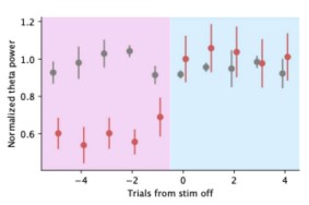
Our stimulation protocol restricted the stimulation to periods that were within the spatial and speed inclusion criteria. The period immediately following the stimulation on every trial is reward delivery, during which the animal has already slowed down, and we do not expect high theta power. Based on the reviewer’s suggestion, we have inspected the first few trials after the stimulus is turned off in the linear track and have confirmed that theta power immediately recovers. We have added these additional figures in Sup. Figure S4H.
Author response image 2.
(4) Do authors have examples of the same trajectory where temporal coding is intact in baseline and disrupted during stimulation? Does an intact theta sequence ever develop in the target animals?
Yes, target animals do exhibit intact spatial sequences during the stimulation-off periods on the linear track and w-track (example in a targeted animal during the stimulation-off condition on linear track below). As we show in Figure 4E and Supplementary Figure S4G, on the w-track, while spatial sequences exist, each cycle’s duration is not consistent across the behavioral experience. Thus, on average, power spectrum of the ahead-behind distance is not rhythmic at 8Hz.
(5) Have the authors computed spike-phase relationships or shown phase-position to evaluate phase precession in individual cells in both the 10 Hz stim vs the phase-specific?
As discussed above, due to unreliable phase estimates during theta suppression, we have chosen to base our analysis of sequential structure on temporal cross-correlation and decoding analysis. However, in response to the reviewer’s question, we have evaluated phase precession under 10Hz stimulation. Consistent with our overall results for the ahead-behind distance, we find that individual cells phase-precess with the newly entrained theta. Additionally, at a population level, we are able to visualize a clear shift in peak spiking frequency.
However, we agree that these results do not completely rule out the contribution of additional spikes, and in a revised version of the manuscript, we have included that possibility.
(6) Did authors perform other types of stimulations that either drove the dominant frequency out of theta range (gamma) or completely desynchronize the system (by stimulating along all different times of theta to perform a phase-specific "scramble")?
We did not attempt a gamma or scrambling stimulation condition.
(7) There is overall inconsistency in the formatting of references throughout the text.
We apologize for these errors and have rectified them in the updated version.



