
Peer review process
Revised: This Reviewed Preprint has been revised by the authors in response to the previous round of peer review; the eLife assessment and the public reviews have been updated where necessary by the editors and peer reviewers.
Read more about eLife’s peer review process.Editors
- Reviewing EditorMohamed TrebakUniversity of Pittsburgh, Pittsburgh, United States of America
- Senior EditorMerritt MadukeStanford University, Stanford, United States of America
Reviewer #1 (Public review):
Summary:
Davis and co-authors used many mouse models to investigate the mechanisms which regulate the contractility of mouse popliteal collecting vessels, primarily chronotropy. The authors use prior literature from the vasculature as a framework to test concepts in lymphatic vessels. The mouse models they used provide evidence for and against the involvement of multiple proteins in regulating chronotropy and other contractile properties in lymphatic vessels. The large amount of data indicate that mechano-stimulation of GNAQ/GNA11-coupled GPCRs generates IP3, which induces intracellular Ca2+ release through IP3R1 to drive depolarization through the activation of ANO1 Cl- channels.
Strengths:
Major strengths of the study are the vast number of mouse knockout models which were used to test the importance of ion channels and G protein signaling pathways in the regulation of lymphatic vessel contractility. The study is a valiant effort and contains a large amount of data. The authors achieved several objectives to find that ANO1 and IP3R1 regulate chronotropy and many other potential proteins do not regulate chronotropy. This study will have a major impact on the field.
Weaknesses:
The authors did an excellent job with the revision. There are no weaknesses.
Reviewer #2 (Public review):
Summary:
In this study, Davis et al., embarked on the quest of the molecular elements responsible for the regulation of lymphatic phasic contractile activity in response to variation of transmural pressure, a mechanism (termed pressure-induced lymphatic chronotropy by the authors). This contractile activity is critical for drainage of interstitial fluid from the tissue and transport of lymph and its cellular and molecular constituents back to the blood circulation. The authors' aim was to investigate the mechanism(s) involved in the pressure-induced regulation of lymphatic pumping, and test whether activation of cation channels, shown in other systems to play mechanosensitive roles are directly at play, and/or whether mechano-activation of Gq/11-coupled GPCRs is necessary to generate second messengers to activate those channels, as it has been suggested for the regulation of myogenic tone in arteries. To achieve their goal, the authors used their well-described, highly reliable protocols of mouse lymphatic vessel isolation, pressure myography, and data acquisition to obtain frequency-pressure relationships and other contractile parameters from transgenic mice where specific channels or molecular elements of interest have been ablated. They combined these data with scRNAseq analysis of these gene targets to determine their respective role and level of expression in lymphatic muscle cells. Their findings demonstrated that, with the exception of ANO1 Cl channels, part of the contractile pacemaker mechanism, none of the exhaustive list of tested ion channels was critical to pressure-induced lymphatic chronotropy, but only ablation of Gq/11 genes or IP3R1 gene in smooth muscle cells led to chronotropy inhibition. To strengthen these findings, they then repeated the myography experiments in the presence of a Gq/11 inhibitor and screened frequency-pressure relationships in vessels from transgenic mice lacking each of the 7 most expressed GPCR according to their scRNAseq dataset. While none of the GPCR ablation showed significant inhibition, they could conclude that transmural pressure activates GPCRs coupled to Gq/11 which generate IP3 to induce SR Ca2+ release through IP3R1 and activate ANO1-mediated depolarization and increase contraction frequency.
Strengths:
The manuscript's strengths reside primarily on very robust, clean and unequivocal pressure myography data and analysis. The research team is mastering these techniques they developed more than a decade ago and have implemented to mouse lymphatics to study their contractile properties, with consistent and convincing outcomes. They also provide data from an impressive list of transgenic mice in order to determine the role of the targeted genes in pressure-induced lymphatic chronotropy, relaying on pharmacological small molecule inhibitors to confirm their findings. Finally, the use of scRNAseq analysis they gathered from previously published dataset brings novelty with respect to expression of the genes of interest in all populations of cells comprising the lymphatic vessels, but more critically to validate or contrast the potential impact of genetic alteration of the given gene in the ability of lymphatic muscles to respond to change in pressure.
Comments on revised version.
The new experiments, additional data, and the now extensive discussion better temper the authors' initial conclusion, the new title as well. The authors should be commended for such an impressive amount of work.
Reviewer #3 (Public review):
In this manuscript, Davis and colleagues aimed to identify the molecular sensors and signaling cascade that enable collecting lymphatic vessels to increase their spontaneous contraction frequency in response to intraluminal pressure (pressure-induced chronotropy). They tested whether the process is similar to blood vessel myogenic constriction by relying on cation channels (TRPC6, TRPM4, PKD2, PIEZO1, etc.) or instead require the activation of G-protein-coupled receptors (presumably mechanosensitive GNAQ/GNA11-coupled receptors), using ex vivo pressure myography of mouse popliteal lymphatics, smooth muscle-specific conditional knockouts, quantitative PCR validation, and single-cell RNA sequencing for target prioritization. The authors convincingly demonstrate that pressure-induced chronotropy does not require the cation channels implicated in arterial myogenic tone but is blunted by deletion of GNAQ/GNA11 or IP3 receptor 1, supporting a model of GPCR > IP3 > Ca2+ release > Cl⁻ channel activation > depolarization. The core conclusion is robust. The work redefines lymphatic pacemaking as G-protein-coupled receptor-dependent mechanotransduction, distinct from arterial mechanisms, and provides a genetically validated toolkit that is useful for studying lymphatic function and dysfunction.
Strengths:
(1) The data are of high quality and highly sensitive functional readouts
(2) The systematic genetic targeting is a major strength that overcomes pharmacological artifacts
(3) Careful quantitative analyses of frequency-pressure slopes
Comments on revised version:
The authors have satisfactorily addressed my comments. No additional comments.
Reviewer #4 (Public review):
Reviewing Editor:
In this revised study Davis and colleagues provide novel findings that are fundamental to the understanding of pressure mechanosensation in lymphatic vessels and are of critical importance to other areas of mechanosensory physiology. The authors used an impressive number of knockout mouse models and employed rigorous state-of-the-art methods, including pressure myography recordings. They provide compelling evidence that mechano-activation of GNAQ/GNA11-coupled GPCRs generates IP3, which induces Ca2+ release from internal stores through IP3R1 and drives depolarization through the activation of ANO1 Cl- channels to induce lymphatic vessel contractility. To uncover the specific GPCR(s) involved in this response, the authors compiled and ranked a list of candidates using single cell RNA sequencing (scRNAseq). In addition to ruling out all major mechanosensitive ion channels previously implicated in this response, the authors also ruled out the involvement of G12/13-coupled GPCRs and 7 most consistently expressed GPCRs in lymphatic smooth muscle along with AT1R. The latter is considered the most likely mechanosensory GPCR in vascular smooth muscle. Although the authors' data fall short of identifying the specific GPCR(s) that mediates pressure-induced chronotropy, they provide compelling evidence that Gq/11-coupled GPCRs are of critical importance to this function. Furthermore, their scRNAseq analysis identified 136 GPCRs in lymphatic smooth muscle, including strongly or moderately expressed candidates such as an olfactory GPCR, three frizzled GPCRs and several adhesion GPCRs (aGPCRs) that encode their own agonist along when many GPCRs with low expression levels. This list represents a foundation for future studies using knockout animal models and aimed at identifying the molecular identity of the GPCR or GPCRs that mediate mechanosensation in lymphatic vessels."
Author response:
The following is the authors’ response to the original reviews.
Public Reviews:
Reviewer #1 (Public review):
Summary:
(1) Davis and co-authors used many mouse models to investigate mechanisms that regulate the contractility of mouse popliteal collecting vessels, primarily chronotropy. Many of the mechanisms studied were previously shown to regulate pressure-induced constriction in small arteries. The authors use prior literature from the vasculature as a framework to test similar concepts in lymphatic vessels. The mouse models used provide evidence for and against the involvement of multiple proteins in regulating chronotropy and other contractile properties in lymphatic vessels. They propose that mechano-activation of GNAQ/GNA11-coupled GPCRs generates IP3, which induces Ca2+ release through IP3R1 and drives depolarization through the activation of ANO1 Cl- channels. Major concerns include the author's major conclusion that GNAQ/GNA11-coupled GPCRs contribute to chronotropy. This conclusion is not supported by the data presented.
Although we have not yet identified a specific GPCR in LMCs that mediates pressure-induced chronotropy, our experiments have established that Gq/11-coupled GPCRs are critical for this response. The almost identical phenotypes of GNAQ/GNA11 double KO vessels with ANO1 KO and IP3R1 KO vessels point to an upstream, mechanosensitive GPCR that regulates ANO1-regulated pacemaking activity through calcium release by IP3R1. New experiments show that the highly specific GNAQ/GNA11 inhibitor YM254890 decreases contraction frequency to the same degree as, or even stronger than, that observed in Gq/11 double KO mice (Fig. 22). Additional data assessing the responses of inguinal-axillary lymphatic vessels (IALVs) from G11-/- and Gq/11 double KO further confirm our findings and conclusions (Suppl. Fig. 1). To address your concern about identifying a specific GPCR, we have performed new scRNAseq analyses to identify and rank the most prominently expressed GPCRs in LMCs (Fig. 21) along with new experiments to test the most likely GPCR candidates that emerged from that analysis (Fig. 22). Please see our specific responses below and the additional data that we now provide.
While our results still fall short of identifying a specific Gq/11-coupled GPCR involved in this LMC mechanosensitive response, they rule out all of the major mechanosensitive ion channels previously implicated in this process (Figs. 4-15), rule out G12/13-coupled GPCRs (Fig.20) and strongly support a critical role for the canonical Gq/11-coupled mechanism (Fig. 18 and Suppl. Fig. 1). New data to eliminate contributions of the 7 most consistently expressed LMC GPCRs, along with the most likely mechanosensory GPCR in VSMCs (AT1R) (Fig. 22), out of the 136 GPCRs detected in our LMC scRNAseq dataset, raise interesting GPCR candidates that require future testing. The remaining list of 11 strongly or moderately expressed candidates includes 3 frizzled GPCRs that typically do not signal through G proteins, an olfactory GPCR, and several adhesion GPCRs (aGPCRs) that encode their own agonist (containing the Stachel peptide sequence). Some of these adhesion GPCRs recruit and signal through Gq/11, and aGPCRs have been implicated in mechanosensation in other cell types (PMID: 25937282, PMID: 40395494). However, pharmacological tools to specifically test most of these aGPCRs are currently lacking. Another possibility, of course, is that the key LMC protein may be a GPCR with a very low expression level.
At present, the only way to test most of those candidates would be to generate new mice with smooth muscle KO of the specific GPCRs, as Offermann’s group has done in a few other cases (PMID: 40623996, PMID: 38597112, PMID: 39920161), but we are without the resources to embark on such an expedition. We are therefore unable to resolve this last issue with currently available resources and/or technology but now acknowledge this as a shortcoming of our study and a future direction. We have adjusted our title to be more congruent with the data presented in the revised manuscript.
Strengths:
(2) One major strength of the study lies in the vast number of mouse knockout models that were used to test the importance of ion channels and G protein signaling pathways in the regulation of lymphatic vessel contractility. In this regard, the study is a valiant effort. The authors achieved several objectives to find that ANO1 and IP3R1 regulate chronotropy, and many other potential proteins do not regulate chronotropy. This study will have a major impact on the field if additional support for G proteins is provided.
Please see our specific responses below and the additional data that we now provide to strengthen support for a GNAQ/GNA11 mechanism.
Weaknesses:
(3) Major conclusions concerning the involvement of G proteins are drawn from the global Gna11 knockout mouse models. This conclusion is weak. Global Gna11 knockout mice are highly likely to have a multifactorial phenotype that could create significant differences in the data.
We agree that global GNAQ/11 DKO mice likely have a multifactorial phenotype and that, IF our hypothesis involved testing some aspect of in vivo function with multiple cell types or organs, this would complicate our conclusions. However, because our approach focuses on ex vivo testing of the F-P relationship of isolated lymphatic vessels, a response mediated specifically by LMCs (i.e., independent of other circulating factors, endothelium, neural control, etc.), our results are NOT likely to be influenced by such extrinsic factors. Stefan Offermanns and colleagues have used similar mice (single floxed gene + Cre + global KO) and ex vivo assays of arteries in multiple peer-reviewed studies (e.g. PMID 31549965), so there is clear precedent for our approach. An alternative approach to the global GNAQ/11 DKO mice would be to use Myh11Cre; GNAQf/f; GNA11f/f double floxed mice, but then those would likely suffer from the potential limitation of competing recombination between the two floxed genes, as we and others have encountered this problem in previous studies using a single Cre with multiple floxed alleles (PMID: 29336844, PMID: 36868507).
(4) Control experiments need to be performed on vessels from the global knockout mice if these major conclusions are to be made.
Control experiments were conducted on popliteal vessels from Myh11Cre; GNAQf/f; GNA11+/+ mice (uninduced littermates) and from GNAQf/f; GNA11+/+ mice (lacking Myh11Cre), the combination of which had a normal F-P relationship (Fig. 18). We have also conducted experiments on Myh11Cre; GNAQf/f; GNA11-/- mice that were not induced with tamoxifen (GNA11-/-). It is not clear if the reviewer is suggesting that we include GNAQ-/- mice, but we did not have access to those mice. Gnaq-/- mice exhibit significant developmental abnormalities (PMID: 22723772), although according to Offermanns and colleagues (PMID: 9687499), only GNAQ homozygous deficient mice displayed “obvious phenotypic defects”.
(5) Similarly, pharmacological tools or alternative approaches to manipulate G proteins should be used to support the data from these mouse models to draw these major conclusions.
To address this comment, we have performed new experiments on WT mice using the potent GNAQ/11 inhibitor YM254890 (at 100 nM, the concentration used in many previous publications by other groups), which had nearly the same effect on abrogating the F-P relationship as induced Myh11Cre; GNAQf/f; GNA11-/- mice (see Fig. 22). The specificity of YM-254890 for the Gq family has recently been verified by excellent work out of David Yule’s lab (PMID: 40563241).
(6) The Gnaq smKO mice are the most specific G protein model studied here. However, there is no phenotype. Do not discuss trends in the data. If the data are not significant, conclude so. If more experiments are required to reach significance, provide more data in the manuscript.
With respect to the Gq smKO data:
(1) We have added more data to the control group.
(2) We have now listed the p value (0.0819 in Fig. 18) for the Gq/11 controls vs Gq smKO, instead of simply designating it “n.s.” with a cut-off of p<0.05; the readers can now judge for themselves how close the values come to being significant.
(3) Although the F-P slope does not reach significance (Fig. 18B), the frequency is significantly different between the Gq/11 controls and Gq smKO at 3 pressures (see the frequency graph in Fig. 18C), so significant differences ARE indeed detected in Gq smKO vessels.
(4) The F-P slope for the DKO is lower than that of the G11 KO and it appears the two knockouts are cumulative, suggesting that Gnaq deletion is contributing to the lower values of the Gq/11 DKO. As further support for this conclusion, we have performed new experiments on IALVs (in part also to address concern #8 below) and that analysis is shown in Suppl. Fig. 1. We include those data as a Supplemental Figure because the contraction data in all the other figures are for popliteal lymphatic vessels. Note that the F-P analysis is not appropriate for IALVs because the control vessels typically show only a 2-2.5x change in frequency over the entire pressure range and frequency essentially reaches a plateau at P = 2 cm H2O. The frequency plots for control, G11-/- and Gq/11 DKO IALV vessels show the same trend as for popliteal vessels (significantly lower than control vessels at all pressures, with only a single exception), but in this case the DKO vessels have significantly lower frequencies than G11-/- vessels at the lower pressures (1-2-3 cm H2O), further supporting the results from popliteal vessels.
(7) The conclusions repeatedly refer to a signaling pathway wherein the upstream component is GPCRs, which activate G proteins. While this may be the case, no GPCRs were identified here, and the involvement of G proteins is questionable, as the authors outline in lines 693-695 and noted above. The conclusions should be tempered, including in the abstract, unless additional experiments are performed to support the involvement of G proteins. Perhaps then the authors may be able to infer that GPCRs are involved.
Please see our response to your comment #1. We have now included additional experiments using the Gq/11 inhibitor YM254890 (at a concentration of 100 nM that is reputed to be “selective” for Gq/11, according to the literature (PMID: 40563241). The blunting of the F-P relationship in the presence of that compound is comparable to, and perhaps even more significant than, the blunting observed for the DKO (the summary analysis is shown for P= 3 cm H2O in the middle of Fig. 22, in a format that matches the other analyses in that figure, but the frequencies were significantly attenuated over the entire lower pressure range). These results strengthen support for a ligand-independent mechanotransduction mechanism through one or more GPCRs of the Gq/11 family. However, because 1) there are at least 136 possible specific GPCR candidates and 2) our new specific tests of the 7 most abundantly expressed GPCRs were negative, pinning down the exact GPCR(s) involved will require much more work. Given the amount of data already presented in this study, and the possibility that the unidentified GPCR is an orphan receptor or other GPCR with very low expression, we think it is appropriate to defer that line of experimentation to a future study. We now clearly state that this is a limitation of the present work and a future direction.
(8) Line 318. The point regarding the choice to use popliteal vessels versus IALVs will be unclear to the uninitiated, particularly as the authors previously used IALVs. Including additional justification in the text and/or data from IALVs in Figure 1, which compares IALVs to popliteal vessels, would better explain the logic.
We now include additional justification in the text by citing Zawieja 2018 and the similarities to Ano1 KO and IP3R1 KO findings in IALVs in our other papers. Note also the very close agreement between the Gq/11 data for popliteal vessels vs IALVs in Fig. 18 C and Suppl. Fig. 1).
(9) The conclusions drawn for TRPC6 and TRPC3 are less convincing. Germline global knockout mice, which are known to undergo compensation, were used, and high data variability is apparent. Using TRPC3 and TRPC6 blockers in the mouse models studied in Figure 4 would strengthen the arguments made regarding these proteins.
Our primary reason for using the global KO mice is that pharmacological inhibitors of TRP channels, particularly TRPC6, are quite non-specific. We did not have access to Trpc6 floxed mice but our ex vivo assay focuses a specific aspect of LMC function in isolation from extrinsic influences (see answer to comment #3 above), and of the 18 cell subtypes detected in these vessels by scRNAseq, Trpc6 is only detectable in LMCs (Fig. 5 panel A). With regard to consequences of global knockouts and possible compensation, that is why we generated the Trpc6/Trpc3 DKO mice, as upregulation of Trpc3 is a known compensatory mechanism for Trpc6 deletion (PMID: 16055711); to our knowledge only one other group has taken the trouble to test this (PMID: 16055711) with respect to vascular smooth muscle function.
(10) Did you perform power analysis to ensure that experimental numbers were sufficient to conclude that no statistical difference exists between datasets? If not, this needs to be done. For example, data shown in Figure 5C for tone and 6C for frequency and tone appear to be significantly different, but are concluded not to be so.
With respect to Fig. 5, there are significant differences in contraction frequency between the TrpC6/C3 DKO mice and their SV129 controls at 5 of 7 pressures, but the frequencies of the KO mice were actually HIGHER—the opposite of the result expected if TRPC6 or TRPC3 were critical for pacemaking. With respect to the effects of TRPC6 KO on tone, there were no significant differences in tone between TRPC6-/- vessels and their SV129 controls, but there were indeed significant differences between the TrpC6/C3 DKO mice and controls at 4 of 7 pressures, but with tone actually being HIGHER than normal (opposite to what would be predicted if those channels were critical for the development of myogenic tone). We point out how that observation is at odds with the conclusions of at least one study on arterial myogenic tone (PMID: 11861411), but that topic is not the focus of our study.
(11) At the end of each result section, a concluding statement is made regarding the effects on pressure-induced chronotropy. In many cases, there are additional effects of manipulating protein expression on other contractile properties. One example is for TRPC3 and TRPC6 (lines 414-416), but others are TRPV4, TRPV3, ENaC, Kir, Cav3.1/3.2, etc. Some interpretation is in the Discussion, but the concluding statements at the end of each result section should be expanded to summarize what the authors think the other significant differences in the data represent.
Thank you for allowing us the flexibility to address this at the end of each relevant section in the Results. We have now expanded several of these sections as you suggest.
(12) Kv7.4 channels. You state you have data (not shown) with linopiridine and XE991. Why not show those results here to support the experiments with the Kcnq4 smKO mice? Otherwise, I suggest you remove the statement from the unpublished data.
We have removed the comments about the unpublished data with other Kv7.4 inhibitors.
(13) Figure 13A. Kcnj2 is modestly expressed in LECs, but very little is present in LMCs. This likely underlies the effect of barium. If you remove the endothelium, does the effect of barium disappear? While this is not the major focus of the study, the effects of barium are dramatic, and it should be made clear whether this is due to inhibition of Kir channels in smooth muscle or endothelial cells.
Thank you for making this very good point. We cannot rule out an effect on LECs in this context and denuding popliteal LVs is nearly impossible without compromising their contractile function because of their small size and the presence of intraluminal valves. The valves prevent easy passage of an air bubble (the opening is only 1/4 the area of the lumen) and attempts at mechanical removal result in extreme valve damage and/or residual LECs at valve sites. If there is an influence of inhibiting Kir channels in LECs, it is not via electrical communication between LECs and LMCs, which is minimal or non-existent as we have shown in previous studies (PMID: 30143234, PMID: 28994159, PMID: 30355030). Thus, a potential LEC influence would be mediated by release/production of vasoactive factors, which is a point that we now discuss.
(14) Figure 18C tone. Several values for losartan look different but are not labelled as such. Please clarify and discuss if different.
All the significantly different values were labeled as such, but that panel is no longer in the revised paper as the AT1R frequency data have been included in a different form (Fig. 22).
(15) The manuscript should include raw data traces in figures that show the major pathways that you conclude regulate chronotropy.
Thank you for this suggestion. Three new figures have now been added to illustrate the blunting of F-P relationship in Ano1 smKO (Fig. 3), IP3R1 smKO (Fig. 17) and GNAQ/11 DKO vessels (Fig. 19).
Reviewer #2 (Public review):
Summary:
In this study, Davis et al. embarked on the quest for the molecular elements responsible for the regulation of lymphatic phasic contractile activity in response to variation of transmural pressure, a mechanism (termed pressure-induced lymphatic chronotropy by the authors) critical for drainage of interstitial fluid from the tissue and transport of lymph back to the blood circulation. Their aim was to investigate the mechanism(s) involved in the pressure-induced regulation of lymphatic pumping, and test whether activation of cation channels, shown in other systems to play mechanosensitive roles are directly at play, and/or whether mechano-activation of GNAQ/GNA11-coupled GPCRs is necessary to generate second messengers to activate those channels, as it has been suggested for the regulation of myogenic tone in arteries. To achieve their goal, the authors used their well-described, highly reliable protocols of mouse lymphatic vessel isolation, pressure myography, and data acquisition to obtain frequency-pressure relationships and other contractile function parameters from transgenic mice where specific channels or molecular elements of interest have been ablated. They combined these data with scRNAseq analysis of these gene targets to determine their respective role and levels of expression in lymphatic muscle cells. Their conclusion is that none of the exhaustive list of tested ion channels was critical, except ANO1 Cl channels, part of the contractile pacemaker mechanism, but that transmural pressure activates GNAQ/GNA11-coupled GPCRs, which generate IP3 to induce SR Ca2+ release through IP3R1 and activate ANO1-mediated depolarization.
Strengths:
The manuscript's strengths reside primarily in very robust, clean, and unequivocal pressure myography data and analysis. The research team is mastering these techniques they developed more than a decade ago and have implemented in mouse lymphatics to study their contractile properties, with consistent and convincing outcomes. They also provide data from an impressive list of transgenic mice in order to determine the role of the targeted gene in pressure-induced lymphatic chronotropy, relying on pharmacological small molecule inhibitors only when necessary. Finally, the use of scRNAseq analysis they gathered from previously published datasets brings novelty with respect to the expression of the genes of interest in all populations of cells comprising the lymphatic vessels, but more critically, to validate or contrast the potential impact of genetic alteration of the given gene on the ability of lymphatic muscles to respond to a change in pressure.
Weaknesses:
(1) The main weakness may reside in the fact that while the authors provide a convincing demonstration that GNAQ/GNA11 are involved in the regulation of the F-P relationship, they give little evidence of the involvement of "upstream" receptors. Indeed, inhibition of AT1R, shown to be involved in myogenic regulation of arteries (a phenomenon the authors rightfully compare to pressure-induced lymphatic chronotropy), didn't lead to a similar effect (decrease in F-P) in lymphatic vessels. Arguably, other GPCRs might be involved in lymphatic vessels, but as such information is not provided in the manuscript, the author's conclusions should be dampened. More in-depth discussion would be required. In fact, it can be argued that the discussion is very restricted with respect to the amount of data and information the manuscript provides.
To address these valid concerns, we performed a detailed scRNA-seq analysis of the GPCRs expressed in mouse LMCs. Approximately 136 GPCRs were identified as being expressed in over 0.5% of LMCs, the top 20 of which are expressed at moderate levels in a substantial percentage (>36%) of LMCs. We have added a new Figure showing bubble plots of these 20 GPCRs in the various cell populations of IALVs, ranked according to their expression level (Fig. 21); we also show their expression in the other cell types. We then experimentally tested 7 of the most likely candidates for which inhibitors were available by determining whether blocking the GPCR would alter pacemaking by lowering the contraction frequency at P=3 cm H2O. This was a streamlined assay compared to measuring the complete F-P relationship but produced similar results; to demonstrate this point, we also performed the streamlined analysis for the ANO1 inhibitor, Ano1 smKO, IP3R1 smKO and Gq/11 DKO, all of which showed significantly reduced frequencies, compared to their respective controls at P=3 cm H2O (see Fig. 22, black symbols). The alignment of statistical significance for the latter results with the corresponding F-P analyses shown in Figs. 2, 16 and 18) demonstrates that this assay is sufficiently powerful to detect a significant effect on pacemaking. Unfortunately, the results for each of the GPCRs tested were negative (Fig. 22, red symbols). We conclude that none of those particular GPCRs (NPY1R, ETAR, NPR3, TBA2XR, S1PR1, AT1AR, AT1BR), or two additional GPCRs (HT-2R, HRH1) that were tested in previous studies (PMID: 12770929, 28453392), are critical for pacemaking or the F-P relationship in LMCs. Of the remaining 11 GPCRs, several are adhesion receptors (Adgrl1, Adgrl2, Adgrl3, Adga2), two are orphan receptors (Gprc5b, Olfr1033), one codes for the GABA-B1 receptor (Gabbr1, with unknown function in the lymphatic system), two code for proteins in the Wnt signaling pathway (Fzd4, Fzd2), one codes for a protein in the Hedgehog signaling pathway (Smo) and one encodes a beta-amyloid binding protein (Tm2d1). The possible roles of these GPCRs, and the other 100+ GPCRs expressed at much lower levels remain to be tested in future studies. At any rate, systematic investigation of these GPCRs is well beyond the scope of this study, and in many cases not possible due the lack of known soluble ligands/antagonists. We have significantly expanded the Discussion with regard to this topic and stated that our inability to identify a specific Gq/11-coupled mechanosensory GPCR in LMCs is a limitation of our study.
(2) Overall, the authors convincingly achieved their aim by performing an impressive number of technically challenging experiments, leading to solid datasets. While these support their main conclusions, a more elaborate discussion might be required to refine them.
We hope this limitation has been tempered by the addition of the new data and the expanded Discussion section mentioned above.
(3) This study is likely to have an important impact on the field as it provides some answers to the lingering question of how lymphatic vessels regulate their contractile activity to variation in transmural pressure and certainly proposes an experimental means to further explore and address that question.
Thank you.
Reviewer #3 (Public review):
In this manuscript, Davis and colleagues aimed to identify the molecular sensors and signaling cascade that enable collecting lymphatic vessels to increase their spontaneous contraction frequency in response to intraluminal pressure (pressure-induced chronotropy). They tested whether the process is similar to blood vessel myogenic constriction by relying on cation channels (TRPC6, TRPM4, PKD2, PIEZO1, etc.) or instead require the activation of G-protein-coupled receptors (presumably mechanosensitive GNAQ/GNA11-coupled receptors), using ex vivo pressure myography of mouse popliteal lymphatics, smooth muscle-specific conditional knockouts, quantitative PCR validation, and single-cell RNA sequencing for target prioritization. The authors convincingly demonstrate that pressure-induced chronotropy does not require the cation channels implicated in arterial myogenic tone but is blunted by deletion of GNAQ/GNA11 or IP3 receptor 1, supporting a model of GPCR > IP3 > Ca2+ release > Cl⁻ channel activation > depolarization. The core conclusion is robust. The work redefines lymphatic pacemaking as G-protein-coupled receptor-dependent mechanotransduction, distinct from arterial mechanisms, and provides a genetically validated toolkit that is useful for studying lymphatic function and dysfunction.
Strengths:
(1) The data are of high quality and highly sensitive functional readouts
(2) The systematic genetic targeting is a major strength that overcomes pharmacological artifacts
(3) Careful quantitative analyses of frequency-pressure slopes
Weaknesses:
(1) The use of inguinal-axillary vessels for single-cell RNA sequencing rather than the popliteal segment studied functionally.
Agreed. The need to use IALVs derives from the much smaller amount of tissue needed for scRNAseq analysis of individual popliteal vessels (and 3-5x longer time required for cleaning) before pooling them. We have tried to clearly state this limitation. However, functional comparisons between popliteal and IALV function are quite similar in almost all aspects that we have studied. In the majority of our protocols, preliminary experiments using IALVs were performed side by side with popliteal vessels (for Ncx1 smKO, Trpc6-/-, Trpc6/3 DKO, G12/13 DKO, Piezo1 smKO, Trmp4 smKO) without detection of a significant difference in phenotype. In addition, we have performed new experiments using IALVs from G11-/- and GNAQ/GNA11 DKO mice to support the major positive findings drawn from the popliteal vessel studies (compare Fig. 18 with Suppl. Fig. 1).
(2) No direct testing of the specific G-protein-coupled receptor involved.
Agreed. As described in our response to the other reviewers (see point 1 to Reviewer 1 and point 1 to Reviewer 2), to address these concerns we have added a new figure (Fig. 21) ranking the top-expressed GPCRs in LMCs and have experimentally tested several of the most likely candidates (Fig. 22). Unfortunately, our results were still negative, allowing us to rule out those particular GPCRs but not to pinpoint a specific GPCR. The possible roles of other 100+ GPCRs in LMCs remain to be investigated in future studies. We have adjusted our title to better reflect our results.
Recommendations for the authors:
Reviewer #1 (Recommendations for the authors):
(1) The authors state multiple times that several cation channels have been shown to contribute to vascular tone. Arterial smooth muscle cell ANO1 channels also contribute to arterial myogenic tone (PMID: 22872152). An ANO1-dependent mechanism is proposed here in lymphatic vessels, and the same ANO1 mouse models are studied. PMID: 22872152 is not discussed or cited and should be.
Now cited.
(2) Lines 88-97. It is important to note that the Bulley paper studied tamoxifen-inducible smooth muscle cell-specific knockout mice, whereas the Sharif-Naeini study used germline SM22-driven PKD1 knockout mice. The mice used in the Sharif-Naeini paper were subsequently found to have an ADPKD-like phenotype due to non-specific knockout of PKD1 in other cell types (PMID: 20856231). That should also be noted. The authors also state "PKD1 channel" in this paragraph when PKD1 does not form a channel.
Corrected and clarified. A new paper on PKD1 by Jaggar’s group is now cited in that context.
(3) Line 119. Perhaps you mean PKD1/2 channels?
Corrected.
(4) Line 312. It is unusual that data from Figure 2A is described before that in Figure 1. Can you reorder the figures to fit the text?
Corrected.
(5) Line 130. Can you provide references that show that ANO1 is not intrinsically mechanosensitive?
Added.
(6) Line 160. I believe the Pkd2f/f mice are available from the Maryland PKD Core and were not generated by the Jaggar lab.
Corrected.
(7) Line 319 needs a reference.
Added.
(8) The color choice for the symbols in many figures, particularly Figures 2, 3, and 4, makes it difficult to know which condition is which.
Corrected.
(9) I may have missed it, but the Figure 1A legend seems to need a definition for every cell type abbreviation.
It is shown in panel A (Figure 2).
(10) In several places, the authors state that they deleted gene expression in LMCs, when the mouse model deletes expression from all smooth muscle cells. Please refine wording.
Clarified.
(11) Line 664-666. Sharif-Naeini did not propose that PKD2 channels contribute to myogenic tone. That was Bulley et al.
Corrected.
Reviewer #2 (Recommendations for the authors):
(1) The role of TRPV4: It is interesting to note that despite a quasi-non-expression of the TRPV4 gene in LMC, KO this gene in these cells results in an increase in contraction frequency and a strong decrease in amplitude at low pressures, suggesting the channel is expressed and active in LMCs. It is not too clear how macrophages could be indirectly involved in this process by releasing products. Are they also sensitive to pressure? How? And why should they be more involved than any other cell type present in the vessel wall (i.e, LECs)?
Macrophages definitely release products that affect LMC activity (PMID: 38826322) and they may be sensitive to pressure but that has not yet been demonstrated. However, as shown in the paper cited, selective KO of Trpv4 in macrophages does not abrogate the F-P relationship in lymphatic vessels.
(2) The role of AT1R in pressure-induced chronotropy: The lack of effect of AT1R inhibition on F-P doesn't mean other GPCRs could play a similar role in lymphatics. A more in-depth discussion could be provided. Furthermore, could G-proteins be activated by pressure independently of coupled receptors?
We have extensively expanded our discussion of possible mechanosensitive GPCRs in LMCs. We have also cited at least one paper providing evidence for G-proteins being activated by pressure independently of coupled receptors (PMID 28148497), although we believe our added assessment of LMC GPCR expression reveals novel targets to explore for mechanotransduction in LMCs.
(3) Figure 1: Please explain the apparent discrepancy between the described protocol and the experiment displayed in A. Please explain and confirm that the experiments presented in the study followed the protocol described in the Methods section. Add text to Methods.
The example does indeed follow the stated protocol in the “METHODS section” for the sequence and duration of pressure steps. Note that we stated 2 min was typical at each pressure level but for some of the lower pressures 4-5 min were required to get a sufficient number of contractions, as in this case. The diameter and pressure traces at P=8, 10 cm H2O are not shown in the example so that we could expand the time axis for better resolution.
It can also be inferred from the trace that the F-P curve was built from frequencies obtained when pressure was lowered and increased. Are values at a given pressure identical if recorded when pressures were lowered or increased? Does it influence the relationship?
There is indeed some variability in the response to stepping pressure up vs down because of rate-sensitive effects, as described in one of our previous studies (PMID: 19001046). In the protocol we implemented here, the FREQ at P=3 cm H2O upon step up from 0.5 cm H2O was usually slightly higher than the initial FREQ at P=3 cm H2O. Those two values were averaged together and this is now stated.
The FREQ graph in B displays values up to 10 cm H2O, which are not shown in A.
The data at P=8, 10 cm H2O were obtained for that same vessel. The complete range is shown to illustrate that the relationship reaches a plateau above 5 cm H2O.
(4) Figure 1: Is there a need to repeat/duplicate the FREQ graph in B and D?
It has now been replaced with Ejection Fraction.
(5) Figure 19: With respect to point #2, and whether G-proteins are involved/important in the process, given the current state of understanding, that part of the illustration might need to be amended.
The figure has been revised.
Reviewer #3 (Recommendations for the authors):
Below are some suggestions for the authors to consider:
(1) Test specific GPCRs pharmacologically (e.g., AT1R) on wild-type vessels to determine if pressure-induced chronotropy is blunted.
Done, Figure 22.
The authors could also rank Gq/11 receptors based on the expression in LMCs (e.g., qPCR using FACS-sorted cells).
Done, Figure 21.
(2) Residual chronotropy in ANO1 knockout mice: Quantify the remaining frequency-pressure slope in Myh11-CreERᵀ²;Ano1ᶠ/ᶠ vessels and test whether it is sensitive to low-chloride buffer or chloride channel blockers to probe an alternative Cl⁻ conductance.
The role of chloride in lymphatic pacemaking is well appreciated and the reviewer offers some intriguing experiments for future experimentation. The use of low Cl- solutions was part of the fundamental discovery of lymphatic muscle pacemaking and the role of a calcium-activated chloride channel as the basis for spontaneous transient depolarizations (STDs) by Dirk Van Helden in 1993 who reported that “Lymphatic STDs reversed at potentials near -35 mV (range -40 to -25 mV; n = 4)” and were “suppressed by exposure to low chloride (replaced with sodium isethionate) solution [(> 5 minutes)]”, although more details were not provided. The effect of low Cl- and lymphatic muscle excitability was followed up in a 2008 paper by von der Weid et al., which showed that STD frequency and amplitude increased acutely (within 2-3 min) after the bath solution was changed to a solution containing 10% of the initial Cl- concentration (replaced with methane sulphonate). This result likely suggests that the acute reduction of extracellular Cl- increased the reversal potential for Cl- and that within 2-3 minutes the intracellular Cl- content had not quite run down. Work by Boedtkjer’s group (Mohanakumar et al. 2018) also demonstrated that human lymphatic pacemaking was also heavily dependent on the bath Cl- concentration. Using wire myography preparations, they showed that human thoracic rings and mesenteric vessels ceased spontaneous contractions when Cl- was removed (replaced with aspartate), and that contractions returned upon washout with a normal Cl- containing solution. In that study, the authors noted that there was some variability in the timing required for contractions to cease with the mesenteric LVs. In some cases, an acute large constriction immediately followed the exchange with Cl- free solution, which would also point to an acute depolarizing/stimulatory effect of a rapid resetting of ECl to a more positive value when external Cl- was removed acutely removed. A limitation to the approach is the use of wire myographs as opposed to pressurized vessels, as the wire method does not allow the vessels to be stretched in a physiological manner. Similarly, diastolic depolarization was not typically observed by Van Helden and von der Weid, as they used pinned-out tissue sections (required for recording STDs from short vessel sections), which highlights the necessity of using a physiological stretch stimulus.
On the face of it, the complete cessation of contractions reported in the Boedtkjer paper is at odds with our previous and present results using Ani9 and Ano1 smKO and IP3R1 smKO vessels, which retain some baseline level of pacemaking, albeit largely pressure-independent. Thus, the hypothesis that a separate Cl- channel may be active and important holds some merit. However, it is worth noting that we do not expect this basal pacemaker drive to be calcium-dependent, as IP3R1 calcium oscillations are maintained. IP3R1 smKO vessels, which largely lack subcellular calcium transients in diastole, also have a lower-than-expected contraction frequency compared to Ano1 smKO vessels, although this is in part due to an elongation of the action potential plateau. We also do not think the residual activity is simply due to the inability of inducible Cres to drive complete recombination, as we performed experiments with Ani9-treated Ano1 smKO vessels (Fig. 2B) and did not observe a significant additional reduction in the concentrations at which maximal inhibition is assumed, 2-10 mM (see also Harlow et al. 2025). We do not see a robust presence of Ano2 (TMEM16b) and while Ano6 (TMEM16) is expressed, it is a calcium-activated scramblase with permeability to both cations and Cl- (Ye et al 2019).
We also have personal experience with Cl- substitution (replaced to 10 mM by aspartate), originally obtained as part of our work identifying Ano1 as the primary calcium-activated chloride channel, although the inability to ascertain the intracellular Cl- at any point in time while the bath exchange is occurring significantly limited the interpretability of that data. During review of that manuscript, we were asked to remove the Cl- substitution experiments but spontaneous contractions still persisted in the majority of the vessels (n=6). Notably this was not a complete removal of Cl- as was done in Mohanakumar et al. 2018. However, the complete removal of Cl- is a much more significant intervention than the inhibition of a single channel, likely altering HCO3- and Na+ flux through the associated Cl- antiporters and symporters. How Cl- removal affects LMC intracellular pH regulation via impaired bicarbonate transport also remains unknown. Nor is it well appreciated how Cl- removal affects mitochondrial function and ROS production in LMCs, which regulate potentially significant confounding pathways related to lymphatic muscle pacemaking. Without control of these significant confounding circumstances, the hypothesis that the baseline depolarization is still a Cl- channel is far from a certainty.
In Author response image 1, we provide the reviewer with the expression data for other members of the Anoctamin family, the potential Anoctamin-regulating CLCA family, and other documented chloride channels in LMCs (clusters 5-6).
Author response image 1.
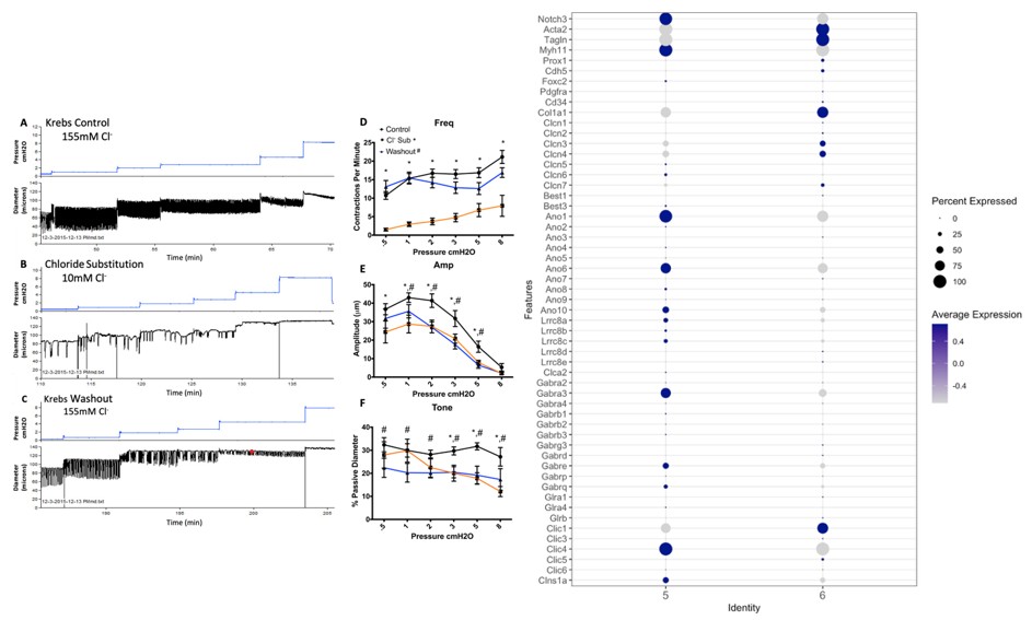
(3) I understand this would be a huge undertaking, but aligning scRNAseq with functional vessels using popliteal LMCs or inguinal-axillary LMCs could help rule out potential tissue-specific differences.
We understand the reviewer’s concerns and, owing to the small amount of cells that can be recovered from popliteal vessels, we instead now provide new data using pressure myography of inguinal axillary vessels (IALVs) for the data with positive findings (Gq/11 inhibition) that support our findings in popliteal vessels (Suppl. Fig. 1). This points to a fundamental role for Gaq, Ga11, and possibly Ga14, in signaling upstream of IP3R1 mobilization and Ano1 activation. We are aware of the length of this manuscript (now further increased in order to respond to the reviewers’ suggestions) and, given the negative data for the vast majority of putative mechanosensory pathways, we have omitted some of the parallel data collected in IALVs. In the majority of our protocols, preliminary experiments using IALVs were tested side by side with popliteal vessels (from Ncx1 smKO, Trpc6-/-, Trpc3/6-/-, G12/13 DKO, Piezo1smKO, Trmp4 smKO mice) without detection of a significant difference in phenotype.



