
Peer review process
Revised: This Reviewed Preprint has been revised by the authors in response to the previous round of peer review; the eLife assessment and the public reviews have been updated where necessary by the editors and peer reviewers.
Read more about eLife’s peer review process.Editors
- Reviewing EditorTomohiro KurosakiThe University of Osaka, Osaka, Japan
- Senior EditorSatyajit RathNational Institute of Immunology, New Delhi, India
Reviewer #1 (Public review):
Summary:
The authors provide in vivo and in vitro evidence for an interaction between AIRE and AID. This has implications for the dynamics of the germinal center response and autoimmunity related to the APSI disease.
Strengths:
Several both biochemical and in vivo experiments to show interaction and the function of AIREs regulation of AID activity in the GC response.
Comments on revised version.
I believe the manuscript is improved.
Reviewer #2 (Public review):
Summary:
In this study, Zhou et al investigated the expression and function of AIRE in B cells in peripheral lymphoid tissues. First, they found the expression of AIRE protein in mature B cells in the follicles in human tonsils and spleens from healthy donors. Flow cytometry analyses using human samples as well as Aire-reporter mice demonstrated AIRE expression in germinal center B cells. The expression of Aire in B cells was induced by CD40 signals. Then, to investigate the impact of AIRE deficiency on B cell function, the authors used a method of transplanting bone marrow cells from Aire-KO and WT mice into B-cell-deficient mice, comparing B cell development and function reconstituted in the recipient mice. Their results showed that Aire-deficient B cells strongly responded to immunization with antigens, exhibiting enhanced class switching and somatic hypermutation of antibodies compared with WT B cells. The same phenomena were observed in CRISPRed B cell lines lacking Aire. The authors successfully utilized the Aire-deficient B cell line to demonstrate that Aire suppresses antibody class switching and somatic hypermutation via its interaction with AID. Finally, using B cell transfer into B cell-deficient mice demonstrated that mice harboring Aire-deficient B cells produced high levels of autoantibodies against Th17 cytokines and exhibited reduced resistance to Candida infection. This mirrors characteristic symptoms in AIRE-deficient patients. The findings of this study not only reveal an unexpected function of AIRE in B cells but also have the potential to contribute to understanding the pathogenesis of APECED and offering a new direction for developing therapies.
Strengths:
The strength of this study lies in demonstrating the expression of function of AIRE in B cells in both mice and humans. It also revealed the direct interaction between AIRE and AID, along with its binding mode (requiring CARD and NLS domains of AIRE), and showed that this interaction is crucial for AIRE function in B cells. It is also significant that the study demonstrated how B cell-intrinsic dysfunction of AIRE leads to autoantibody production against cytokines.
Comments on revised version.
My previous concerns have been properly addressed.
Author response:
The following is the authors’ response to the original reviews.
eLife Assessment
AIRE has been well known to contribute to immune self-tolerance in the thymus by expressing auto-antigens; in this manuscript, the authors describe unexpected findings about the interaction of AIRE with AID in B cells, and its function in the immune system, thereby contributing to a fundamental understanding of the broader functions of AIRE. The strength of this manuscript is that, by employing biochemical and genetic experiments, the authors convincingly show interaction between AIRE and AID and subsequent AIRE's function in the GC responses. However, two weak points exist: first, the connection between AIRE, auto-anti IL17 Abs, and IL17-positive effector T cells, and second, like the thymus, expression of auto-antigens by AIRE in the GC B cells has not been tested.
We thank the journal editors for the thoughtful and constructive evaluation of our data and for recognizing the significance, strengths and weaknesses of our study. We have addressed these major weak points in the responses below and have also provided discussion on the important aspect of whether AIRE regulates autoantigen expression in GC B cells analogous to its role in mTECs, which certainly warrants future investigation.
Public Reviews:
Reviewer #1 (Public review):
Summary:
The authors provide in vivo and in vitro evidence for an interaction between AIRE and AID. This has implications for the dynamics of the germinal center response and autoimmunity related to the APSI disease.
The manuscript describes an unexpected function of AIRE, which is more well known for its function to regulate negative selection of T cells in the thymus. Here, the gene has also been shown to be expressed by B cells (Immunity 2015: 26070482). They describe that AIRE interacts with AID, and in its absence, B cells acquire more hypermutations and also produce autoantibodies against IL-17. These autoantibodies have been described previously.
Strengths:
The study is interesting and provides some additional information about how AIRE regulates immune cell function. Several biochemical and in vivo experiments show the interaction and the function of AIREs in the regulation of AID activity in the GC response.
Weaknesses:
Some of the hypothetical consequences of this regulation are not investigated. This includes responses to model antigens and dynamics of the germinal center related to kinetics.
We are grateful for the reviewer’s interest and careful and thoughtful review of the manuscript.
Major Comments:
(1) AID regulates both switch and somatic hypermutation. Switch is easier to achieve, so which of these processes does AIRE influence the most? Also, the switch is thought to occur before the B cell enters the GC. Looking at the histology, is AIRE also expressed at the early proliferative stage that has been described by Ann Haberman?
We thank the reviewer for raising this provocative point. Yes, it has been recently shown that CSR occurs earlier after B cell activation largely prior to GC entry (Roco et al. 2019), and CSR is generally easier to induce in vitro than SHM. These data and observations further support well-established findings that CSR and SHM involve distinct mechanisms and pathways to resolve DNA lesions (Frossi et al., 2019; Masani et al., 2013; Roco et al., 2019; Schrader et al., 2023), even though both require AID. We observed a strong upregulation of AIRE expression upon CD40 signaling (Fig. 2C–G) and that AIRE in B cells negatively regulates both CSR and SHM (Fig. 3E– J and Fig. 4A–H), but it would be difficult to directly compare the magnitude of AIRE’s influence between them because they are mechanistically distinct and occurs at different stages and locations during antigen-specific B cell responses.
We observed some AIRE-expressing B cells outside of the GC (Fig. 1A–C, J), some of which could represent newly activated cells about to enter the GC reaction, but we did not perform imaging or flow cytometry experiments or track their Ki67 or Bcl6 expression in the AireAdig reporter mice. However, these cells appear to be largely IgD-, suggesting that they may have already undergone CSR and are perhaps not (entirely) the early proliferative B cells described by Dr Haberman.
(2) In experiments determining anti-CD40-dependent upregulation of AIRE, naïve resting B cells were used from mice. A proportion of the B-cells got activated. Are these MZB or FOB cells as MZBs are more easily activated?
We thank the reviewer for this careful interpretation of our data. Indeed, the methods used to purify naïve B cells do not exclude MZBs and, as they are more sensitive to activation, may also express AIRE upon stimulation with CD40L. However, although we cannot exclude MZBs in these cultures, they represent less than 10% of the total B cells in these cultures whereas we observed approximately 30% of the cells upregulating AIRE expression.
Interestingly, Yamano et al. (Yamano et al., 2015) suggested that GC B cells may not express AIRE due to strong BCR signaling; however, it is known that GC B cells have attenuated BCR signaling to promote LZ to DZ transition, consistent with the timing of AIRE upregulation postCD40-CD40L engagement (Davidzohn et al., 2020; Khalil et al., 2012). In contrast, MZBs are known to exhibit greater baseline activation of pathways downstream of BCRs (Hampel et al., 2011), which may provide inhibitory signals for the upregulation of AIRE.
(3) In the BM chimeric experiments in Figure 3. Do the AIRE+ and AIRE - populations distribute equally among B cell subpopulations?
We thank the reviewer for this interesting comment. In our chimera experiment, although we did not analyze specific subsets such as B-1 or MZB, we observe that AIRE-deficient B cells outcompeted WT B cells in lymphoid organs as well as in the blood in spite of prior publications showing equivalent reconstitution of CD45.2 mice with CD45.1/CD45.2 B cells (Kalari Kandy et al., 2023).
(4) Furthermore, in the NP-KLH experiments, one would expect that B cells with increased affinity would leave the GC earlier and become plasma cells. Thus, the kinetics of the AIRE+ vs AIRE- B cells within the GC would be different? Also, would they maybe take over at some point, as the increased affinity would favor help from Tfh cells that are known to be limited?
We thank the reviewer for these insightful comments. Yes, we would expect to see that Aire-/- B cells will dominate GCs over time. Indeed, our chimera experiments (Fig. 3A–D) indicated that Aire-/- B cells were of higher frequency in the GCs compared to WT post-immunization, consistent with the expansion of higher affinity clones. However, although we did not quantify the development of PCs in these mice, our adoptive transfer experiments showed that mice receiving Aire-/- B cells developed higher affinity antibodies compared to those receiving WT B cells (Fig. 3G), indicating a higher affinity plasma cell pool compared to controls.
(5) Given the previous studies on AIRE's function in regulating transcription (PMID: 34518235), how does this interaction fit into this picture?
We thank the reviewer for raising this important point. Although we did not directly test the role of AIRE in the regulation of tissue-restricted antigens, we observed a clear interaction between AIRE and pSer5 Pol II (Fig. 6F), consistent with its function as a broad-spectrum transcriptional regulator (Fang et al., 2024; Giraud et al., 2012; Oven et al., 2007). This is particularly important for antibody diversification in B cells, as it is known that AID is targeted to sites of Pol II pausing (Chaudhuri et al., 2003; Pavri et al., 2010). Further, recent reports have shown that the CARD domain of AIRE promotes its polymerization and the formation of nucleation sites at which a positive feedback loop to create transcriptional hubs (Huoh et al., 2024). Interestingly, the CARD domain is also necessary for the interaction between AIRE and AID (Fig. 5F), indicating that either these condensates are also critical for preventing AID from being recruited to Pol II or that AIRE may perform alternative functions in B cells compared to mTECs. Nevertheless, in this current manuscript, we focus on the capacity of AIRE to utilize this interaction with pSer5 Pol II to prevent AID localization to its DNA substrates, however, future studies may focus on how this interaction may impact the expression of peripheral tissue antigens to promote T cell tolerance.
(6) In the uracil experiments, the readout for AID to induce double-stranded breaks could be tested.
We thank the reviewer for this suggestion for complementary data to our uracil analyses. In our manuscript, we tested the generation of uracil in Aire+/+ and Aire-/- CH12 cells since this is a downstream function of AID’s activity. Therefore, our data focused on an immediate and direct impact of AIRE on AID’s activity. Further, although we observe an interaction between AIRE and AID as well as a downstream functional consequence, it is unclear whether AIRE may also impact other pathways in these cells after activation that may confound the results of downstream analyses, such as DSBs. We agree that DSBs would be an interesting readout further downstream, and future work may focus on the function of AIRE in these additional processes.
(7) The candida experiments are a nice connection to the situation in patients. However, why is it mostly auto-antibodies against IL-17? How about other immune responses, as well as T cellindependent type I and II responses?
We thank the reviewer for raising this important point. Indeed, we observed a significant increase in the generation of autoreactive, neutralizing antibodies against Th17-associated cytokines (Fig. 7D, E). However, although AIRE-deficient patients produce neutralizing antibodies against type 1 interferons as well, clearance of the fungal pathogen Candida albicans relies heavily on IL-17 to promote the upregulation of antimicrobial peptides and neutrophil infiltration (Conti et al., 2014), which may be a particularly pronounced response induced in our mouse models. Therefore, we targeted our assay towards these cytokines, but do not rule out possible production of autoantibodies against other factors. It would be interesting for additional studies to determine the production of autoreactive antibodies against cytokines in other infection models, such as viral infections, as APS-1 patients have been reported to display an increased susceptibility to viral infections as well (Bastard et al., 2021; Hetemaki et al., 2021; Oikonomou et al., 2021).
These important discussions have been included in the revised manuscript.
Reviewer #2 (Public review):
Summary:
In this study, Zhou et al investigated the expression and function of AIRE in B cells in peripheral lymphoid tissues. First, they found the expression of AIRE protein in mature B cells in the follicles in human tonsils and spleens from healthy donors. Flow cytometry analyses using human samples as well as Aire-reporter mice demonstrated AIRE expression in germinal center B cells. The expression of Aire in B cells was induced by CD40 signals. Then, to investigate the impact of AIRE deficiency on B-cell function, the authors used a method of transplanting bone marrow cells from Aire-KO and WT mice into B-cell-deficient mice, comparing B-cell development and function reconstituted in the recipient mice. Their results showed that Aire-deficient B cells strongly responded to immunization with antigens, exhibiting enhanced class switching and somatic hypermutation of antibodies compared with WT B cells. The same phenomena were observed in CRISPRed B cell lines lacking Aire. The authors successfully utilized the Aire-deficient B cell line to demonstrate that Aire suppresses antibody class switching and somatic hypermutation via its interaction with AID. Finally, using B cell transfer into B cell-deficient mice demonstrated that mice harboring Aire-deficient B cells produced high levels of autoantibodies against Th17 cytokines and exhibited reduced resistance to Candida infection. This mirrors characteristic symptoms in AIRE-deficient patients. The findings of this study not only reveal an unexpected function of AIRE in B cells but also have the potential to contribute to understanding the pathogenesis of APECED and to offering a new direction for developing therapies.
We are grateful for the reviewer’s careful and thoughtful review of the manuscript and appreciation of the significance of our findings.
Strengths:
The strength of this study lies in demonstrating the expression of the function of AIRE in B cells in both mice and humans. It also revealed the direct interaction between AIRE and AID, along with its binding mode (requiring CARD and NLS domains of AIRE), and showed that this interaction is crucial for AIRE function in B cells. It is also significant that the study demonstrated how B-cell-intrinsic dysfunction of AIRE leads to autoantibody production against cytokines.
Weaknesses:
As for loss-of-function analysis of Aire in B cells, in addition to the B cell transfer from Aire-KO mice performed in this study, generating B cell-specific Aire-deficient mice using Aire-flox mice (Dobes et al, Eur J Immunol 2018) would further reinforce the conclusions of this study. Furthermore, the relationship with Aire function in thymic B cells reported by previous studies remains unclear, posing an unresolved challenge. This study also failed to address whether Aire deficiency affects gene expression in GC B cells, in particular, whether it induces the expression of various self-antigens as reported in thymic B cells or mTECs.
We thank the reviewer for these thoughtful critiques of our manuscript. As this study was largely carried out prior to the development of Aire-flox mice, we took advantage of the adoptive transfer model to generate mice with AIRE deletion specifically in B cells. Since Aire-flox mice were developed, these mice would be the new gold standard for analysis and would provide further strength to our existing data. Unfortunately, these mice are not readily available, and we do not currently have the resources to reconstitute this line and are unable to obtain them.
In addition, although we do not directly test the role of AIRE in the regulation of tissue-restricted antigens, we observed a clear interaction between AIRE and pSer5 Pol II (Fig. 6F), consistent with its function as a transcriptional regulator (Fang et al., 2024; Giraud et al., 2012; Oven et al., 2007). This is particularly important for antibody diversification in B cells, as it is known that AID is targeted to sites of Pol II pausing (Chaudhuri et al., 2003; Pavri et al., 2010). Further, recent reports have shown that the CARD domain of AIRE promotes its polymerization and the formation of nucleation sites at which a positive feedback loop creates transcriptional hubs (Huoh et al., 2024). Interestingly, the CARD domain is also necessary for the interaction between AIRE and AID (Fig. 5F) indicating that either these condensates are also critical for preventing AID from being recruited to Pol II or that AIRE may perform alternative functions in this subset of B cells compared to mTECs. Nevertheless, in this study, we focused on the capacity of AIRE to utilize this interaction with pSer5 Pol II to reduce AID targeting to its DNA substrates. However, future studies may focus on how this interaction may impact the expression of peripheral tissue antigens to promote T cell tolerance.
The discussion related to these important aspects raised by the reviewer are included in the original and revised manuscript.
Recommendations for the authors:
Reviewer #1 (Recommendations for the authors):
(1) How do these findings connect to IgA responses and the ongoing GC response in the peyers patches?
We thank the reviewer for raising this additional point. Indeed, we also observed AIRE expression in GC B cells from Peyer’s patches (Fig. S1J) and found that CSR to IgA in our in vitro models was increased by the deletion of AIRE in B cells (Fig. 4A–C). These data suggest that, in AIRE-deficient B cells, there may be an increase in IgA production, however, we did not directly test mucosal IgA levels in our adoptive transfer models. Future work may focus on the role of B cell intrinsic AIRE and its impact on the capacity of Peyer’s patch-associated B cells to produce IgA and its role on mucosal barrier immunity and microbiota coating.
These important discussions have been included in the revised manuscript.
(2) Some of the histology is rather dark, and it would be nice to see some more examples in the supplement.
We thank the reviewer for bringing this to our attention. We have included an additional staining of a whole follicle with GC in the supplement as requested (Fig. S1C, bottom panel).
Reviewer #2 (Recommendations for the authors):
(1) In the Discussion section, could the authors explore Aire's functions and biological significance in greater depth? Although Aire was originally identified as a regulator of tissue-restricted antigen expression in mTECs, it has subsequently shown to play crucial roles in diverse processes, including germ cell development, antigen processing, and B cell function regulation (this study). In light of these recent findings, it is conceivable that Aire did not evolve specifically as an mTECassociated gene. Rather, the reverse may be possible: Aire may have originally emerged as a regulator of germ cell development and/or immune responses, with its expression and functions later co-opted by mTECs (analogous to how other transcription factors have been adopted for differentiation of mimetic mTECs). I would welcome the authors expanding on this evolutionary perspective in future work or revisions.
We thank the reviewer for this insightful and fascinating perspective on the evolutionary and ancestral functions of AIRE as a transcription factor. We have incorporated additional discussion in the revised manuscript.
(2) Personally, I knew that Aire is expressed in B cells, as indicated by several gene expression databases. But the ImmGen database shows that Aire mRNA expression is detected at higher levels in plasma cells than in GC B cells. What do the authors think?
We sincerely thank the reviewer for this question and appreciate the personal comment in support of our results. Indeed, the ImmGen database shows little Aire expression in GC B cells, with most of the signal coming from plasma cells in the B cell compartment. Further, as the Reviewer suggested, reanalysis of previously published scRNA-seq (Duan et al., 2021) has shown Aire transcript in some, albeit a lower percentage of, GC B cells (Author response image 1), which was more abundant near the peak time of the immune response after immunization, in line with our and the Reviewer’s observations.
Author response image 1.
Analysis of Aire expression in the day 7 (upper row) and day 14 (lower row) post-immunization scRNA-Seq datasets (Duan et al., 2021), showing 200 Aire+ GC B cells out of 3542 GC B cells (5.65%) on day 7 and 183 Aire+ GC B cells out of 19757 GC B cells (0.93%) on day 14, and the distribution of these Aire+ GC B cells in both dark zone (DZ) and light zone (LZ) B cells.
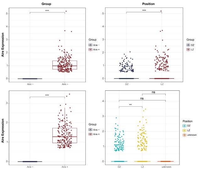
A myriad of technical factors can impact the detection of lower-expressed genes by bulk RNA-seq and even scRNA-seq, and that looking at individual cells may give better resolution. This is the case for a considerable number of other genes we have studied or that have been reported in other investigators’ publications. Many biological factors can also have an important effect (e.g. protein levels may vary depending on cell states and do not necessarily correlate with transcript levels). Significant impact of the strain background, diet, microbiome and the living environment of the animals cannot be excluded either. Similarly, recent reports have shown that CGRP from spleen-innervating nociceptors signals to splenic B cells to promote germinal center reactions (Wu et al., 2024). However, Immgen showed no Ramp1 expression in splenic B cell compartments. In addition, Nur77/Nr4a1 has also been shown to be highly expressed by a small number of light zone cells and is critical for regulating GC reactions (Brooks et al., 2021; Mueller et al., 2015) even though ImmGen shows very little expression of this gene in its bulk sequencing. Therefore, we think that much of the data from ImmGen should be carefully validated with additional experimental methods. In this case, we were able to detect AIRE protein using methods including immunofluorescence, immunoprecipitation, western blot, and flow cytometry.
(3) Line 343: "donor Aire-/-" may be a mistake for "donor Aire+/+"?
We thank the reviewer for bringing this to our attention and have adjusted the manuscript accordingly.
(4) Line 346: Is "data now shown" a spelling mistake? Including this, there are multiple occurrences of "data not shown" in the Results and Discussion sections. If the authors describe or discuss the data, it should be shown.
We thank the reviewer for bringing this to our attention and have revised the manuscript accordingly.
(5) Line 346-350: The authors should investigate more thoroughly whether Aire influences gene expression in GC B cells, particularly the expression of a set of self-antigen genes, as demonstrated in thymic B cells by Yamano et al (Immunity 2015). Regardless of the outcome, such data would be highly significant for this study and the broader research community.
We thank the reviewer for raising this important point. Although we did not directly test the role of AIRE in the regulation of tissue-restricted antigens, we observed a clear interaction between AIRE and pSer5 Pol II (Fig. 6F), consistent with its function as a transcriptional regulator (Fang et al., 2024; Giraud et al., 2012; Oven et al., 2007). This is particularly important for antibody diversification in B cells, as it is known that AID is targeted to sites of Pol II pausing (Chaudhuri et al., 2003; Pavri et al., 2010). Further, recent reports have shown that the CARD domain of AIRE promotes its polymerization and the formation of nucleation sites at which a positive feedback loop to create transcriptional hubs (Huoh et al., 2024). Interestingly, the CARD domain is also necessary for the interaction between AIRE and AID (Fig. 5F) indicating that either these condensates are also critical for preventing AID from being recruited to Pol II or that AIRE may perform alternative functions in B cells compared to mTECs. Nevertheless, in this current study, we focused on the capacity of AIRE to utilize this interaction with pSer5 Pol II to prevent AID targeting to its DNA substrates, and future studies may focus on how this interaction may impact the expression of peripheral tissue antigens to promote T cell tolerance.
(6) Line 1282-1293: The number of B cells used for Western blotting should be clearly indicated. Since the expression levels of Aire mRNA in B cells are lower than those in mTECs, detecting Aire protein in B cells is also likely difficult. It is important to specify the number of B cells required so that other researchers can reproduce the Western blotting results presented in this paper.
We thank the reviewer for raising this important point and have revised the manuscript accordingly.
References
Bastard, P., E. Orlova, L. Sozaeva, R. Levy, A. James, M.M. Schmitt, S. Ochoa, M. Kareva, Y. Rodina, A. Gervais, T. Le Voyer, J. Rosain, Q. Philippot, A.L. Neehus, E. Shaw, M. Migaud, L. Bizien, O. Ekwall, S. Berg, G. Beccuti, L. Ghizzoni, G. Thiriez, A. Pavot, C. Goujard, M.L. Fremond, E. Carter, A. Rothenbuhler, A. Linglart, B. Mignot, A. Comte, N. Cheikh, O. Hermine, L. Breivik, E.S. Husebye, S. Humbert, P. Rohrlich, A. Coaquette, F. Vuoto, K. Faure, N. Mahlaoui, P. Kotnik, T. Battelino, K. Trebusak Podkrajsek, K. Kisand, E.M.N. Ferre, T. DiMaggio, L.B. Rosen, P.D. Burbelo, M. McIntyre, N.Y. Kann, A. Shcherbina, M. Pavlova, A. Kolodkina, S.M. Holland, S.Y. Zhang, Y.J. Crow, L.D. Notarangelo, H.C. Su, L. Abel, M.S. Anderson, E. Jouanguy, B. Neven, A. Puel, J.L. Casanova, and M.S. Lionakis. 2021. Preexisting autoantibodies to type I IFNs underlie critical COVID-19 pneumonia in patients with APS-1. The Journal of experimental medicine 218:
Brooks, J.F., C. Tan, J.L. Mueller, K. Hibiya, R. Hiwa, V. Vykunta, and J. Zikherman. 2021. Negative feedback by NUR77/Nr4a1 restrains B cell clonal dominance during early Tdependent immune responses. Cell Rep 36:109645.
Chaudhuri, J., M. Tian, C. Khuong, K. Chua, E. Pinaud, and F.W. Alt. 2003. Transcription-targeted DNA deamination by the AID antibody diversification enzyme. Nature 422:726-730.
Conti, H.R., A.R. Huppler, N. Whibley, and S.L. Gaffen. 2014. Animal models for candidiasis. Curr Protoc Immunol 105:19.16.11-19.16.13.
Davidzohn, N., A. Biram, L. Stoler-Barak, A. Grenov, B. Dassa, and Z. Shulman. 2020. Syk degradation restrains plasma cell formation and promotes zonal transitions in germinal centers. The Journal of experimental medicine 217:
Duan, L., D. Liu, H. Chen, M.A. Mintz, M.Y. Chou, D.I. Kotov, Y. Xu, J. An, B.J. Laidlaw, and J.G. Cyster. 2021. Follicular dendritic cells restrict interleukin-4 availability in germinal centers and foster memory B cell generation. Immunity 54:2256-2272 e2256.
Fang, Y., K. Bansal, S. Mostafavi, C. Benoist, and D. Mathis. 2024. AIRE relies on Z-DNA to flag gene targets for thymic T cell tolerization. Nature 628:400-407.
Frossi, B., G. Antoniali, K. Yu, N. Akhtar, M.H. Kaplan, M.R. Kelley, G. Tell, and C.E.M. Pucillo. 2019. Endonuclease and redox activities of human apurinic/apyrimidinic endonuclease 1 have distinctive and essential functions in IgA class switch recombination. J Biol Chem 294:5198-5207.
Giraud, M., H. Yoshida, J. Abramson, P.B. Rahl, R.A. Young, D. Mathis, and C. Benoist. 2012. Aire unleashes stalled RNA polymerase to induce ectopic gene expression in thymic epithelial cells. Proceedings of the National Academy of Sciences of the United States of America 109:535-540.
Hampel, F., S. Ehrenberg, C. Hojer, A. Draeseke, G. Marschall-Schroter, R. Kuhn, B. Mack, O. Gires, C.J. Vahl, M. Schmidt-Supprian, L.J. Strobl, and U. Zimber-Strobl. 2011. CD19independent instruction of murine marginal zone B-cell development by constitutive Notch2 signaling. Blood 118:6321-6331.
Hetemaki, I., S. Laakso, H. Valimaa, I. Kleino, E. Kekalainen, O. Makitie, and T.P. Arstila. 2021. Patients with autoimmune polyendocrine syndrome type 1 have an increased susceptibility to severe herpesvirus infections. Clin Immunol 231:108851.
Huoh, Y.S., Q. Zhang, R. Torner, S.C. Baca, H. Arthanari, and S. Hur. 2024. Mechanism for controlled assembly of transcriptional condensates by Aire. Nature immunology 25:15801592.
Kalari Kandy, R.R., X. Fan, and X. Cao. 2023. CD45.1/CD45.2 Congenic Markers Induce a Selective Bias for CD8+ T Cells during Adoptive Lymphocyte Reconstitution in Lymphocytopenia Mice. Immunohorizons 7:755-759.
Khalil, A.M., J.C. Cambier, and M.J. Shlomchik. 2012. B cell receptor signal transduction in the GC is short-circuited by high phosphatase activity. Science 336:1178-1181.
Masani, S., L. Han, and K. Yu. 2013. Apurinic/apyrimidinic endonuclease 1 is the essential nuclease during immunoglobulin class switch recombination. Mol Cell Biol 33:1468-1473.
Mueller, J., M. Matloubian, and J. Zikherman. 2015. Cutting edge: An in vivo reporter reveals active B cell receptor signaling in the germinal center. Journal of immunology 194:29932997.
Oikonomou, V., T.J. Break, S.L. Gaffen, N.M. Moutsopoulos, and M.S. Lionakis. 2021. Infections in the monogenic autoimmune syndrome APECED. Curr Opin Immunol 72:286-297.
Oven, I., N. Brdickova, J. Kohoutek, T. Vaupotic, M. Narat, and B.M. Peterlin. 2007. AIRE recruits P-TEFb for transcriptional elongation of target genes in medullary thymic epithelial cells. Mol Cell Biol 27:8815-8823.
Pavri, R., A. Gazumyan, M. Jankovic, M. Di Virgilio, I. Klein, C. Ansarah-Sobrinho, W. Resch, A. Yamane, B. Reina San-Martin, V. Barreto, T.J. Nieland, D.E. Root, R. Casellas, and M.C. Nussenzweig. 2010. Activation-induced cytidine deaminase targets DNA at sites of RNA polymerase II stalling by interaction with Spt5. Cell 143:122-133.
Roco, J.A., L. Mesin, S.C. Binder, C. Nefzger, P. Gonzalez-Figueroa, P.F. Canete, J. Ellyard, Q. Shen, P.A. Robert, J. Cappello, H. Vohra, Y. Zhang, C.R. Nowosad, A. Schiepers, L.M. Corcoran, K.M. Toellner, J.M. Polo, M. Meyer-Hermann, G.D. Victora, and C.G. Vinuesa. 2019. Class-Switch Recombination Occurs Infrequently in Germinal Centers. Immunity 51:337-350 e337.
Schrader, C.E., T. Williams, K. Pechhold, E.K. Linehan, D. Tsuchimoto, and Y. Nakabeppu. 2023. APE2 Promotes AID-Dependent Somatic Hypermutation in Primary B Cell Cultures That Is Suppressed by APE1. Journal of immunology 210:1804-1814.
Wu, M., G. Song, J. Li, Z. Song, B. Zhao, L. Liang, W. Li, H. Hu, H. Tu, S. Li, P. Li, B. Zhang, W. Wang, Y. Zhang, W. Zhang, W. Zheng, J. Wang, Y. Wen, K. Wang, A. Li, T. Zhou, Y. Zhang, and H. Li. 2024. Innervation of nociceptor neurons in the spleen promotes germinal center responses and humoral immunity. Cell 187:2935-2951 e2919.
Yamano, T., J. Nedjic, M. Hinterberger, M. Steinert, S. Koser, S. Pinto, N. Gerdes, E. Lutgens, N. Ishimaru, M. Busslinger, B. Brors, B. Kyewski, and L. Klein. 2015. Thymic B Cells Are Licensed to Present Self Antigens for Central T Cell Tolerance Induction. Immunity 42:1048-1061.



