
Peer review process
Revised: This Reviewed Preprint has been revised by the authors in response to the previous round of peer review; the eLife assessment and the public reviews have been updated where necessary by the editors and peer reviewers.
Read more about eLife’s peer review process.Editors
- Reviewing EditorPatrick ForcelliGeorgetown University, Washington, United States of America
- Senior EditorJohn HuguenardStanford University School of Medicine, Stanford, United States of America
Reviewer #1 (Public review):
Summary:
The manuscript entitled "Autonomic reflex plasticity associates with time-dependent SUDEP susceptibility in a murine model with hyperactive stress circuits" by Dr. Saunders and colleagues combined a traditional mouse model of SUDEP, ventral intrahippocampal kainite (vIHKA) injection, with a genetic model of chronic hyperactivity of central corticotropin-releasing hormone (CRH) neurons (Kcc2/Crh) that further increases the risk of SUDEP in the weeks following seizure.
Strengths:
Their results show during spontaneous seizures Kcc2/Crh mice had more pronounced reflex-like ictal bradycardias compared to WT controls that notably occurred prior (~10 sec) to seizure termination and had greater autonomic disturbances compared to WT controls, including a pronounced serotonin-mediated Bezold Jarisch reflex. These results show chronic hyperactivity of central corticotropin-releasing hormone (CRH) neurons (Kcc2/Crh) increased autonomic disturbances and risk of SUDEP in a kainic acid model of epilepsy.
Weaknesses:
This study could be improved with a more thorough assessment of heart rate, blood pressure and breathing during and following the seizures, and in particular the fatal event. It is unclear if the bradycardias were spontaneous, or a result of preceding central or obstructive apneas, oxygen desaturations, hypercapnia, arrhythmias, or other possible triggers.
Considerable prior work in the literature suggests SUDEP could be mediated, in some patients, by a burst of parasympathetic activity to the heart. Were the heart rate changes in these animals during seizures inhibited or blocked by atropine, or atenolol? The injection of the 5HT agonist phenylbiguanide into the right jugular is not a selective approach for activating the Bezold Jarisch Reflex (BJR) which is caused by increased activity of intracardiac sensory neurons (generally activated with ischemia or a combination of low preload with high contractility). The results should be interpreted more cautiously, as a response to systemic administration of phenylbiguanide only.
Reviewer #2 (Public review):
Summary:
In this manuscript, the authors set out to evaluate the role of hypothalamic pituitary axis hyperactivity on cardiac and autonomic changes during epileptogenesis and following seizures in a mouse model of temporal lobe epilepsy. Epilepsy is very common. It can frequently result in death from sudden unexpected death in epilepsy, or SUDEP. SUDEP is thought to be at least in part due to seizure related cardiac and autonomic instability. Increased stress states are well known to be comorbid with epilepsy. This comorbidity is thought to increase the risk of SUDEP. Here the authors hypothesized that a mouse model of heightened stress in which there is hyperactivity of the CRH neurons in the hypothalamus would demonstrate exaggerated cardiac and autonomic effects of seizures and epilepsy.
Strengths:
For the chronic stress model, they employed the Kcc2/Crh mice that have a genetic deletion of the potassium chloride cotransporter in CRH neurons. They treated these mice and their wild type littermates with intra hippocampal kainic acid or saline, as epileptic and sham-treated animals respectively. The assessed cardiac activity, blood pressure, baroreflex, and the Bezold-Jerisch reflex during epileptogenesis. This in general is an interesting study. They make some interesting and potentially important observations regarding heart rate and blood pressure in seizures and epilepsy.
Weaknesses:
While the revised manuscript is much improved, there are still some concerns that should be addressed.
(1) The low-pressure baroreceptor responses they show in Figure 4 are somewhat confusing. Should they not be seeing a reflex increase in heart rate when blood pressure is lowered with sodium nitroprusside? It would be helpful if they could describe whether the control responses were as expected or not, and if not, why? This makes it difficult to assess changes seen in the different genotypes and conditions.
(2) It does not seem appropriate to label the assessments associated with Figure 5 as the Bezold Jarisch Reflex. This reflex involves bradycardia, hypotension, vasoconstriction, and hypopnea. They seem to be only looking at the cardiac component, which is likely mediated though peripheral 5HT3 receptors. Did they measure blood pressure and breathing? Can they include these? If they can only comment on HR, then the discussion should reflect this.
(3) In Figure 1B, it would be helpful to show some short (e.g., 0.5 sec) snippets of ECG traces that exemplify the changes in HR (spikes/second).
(4) The day 21 examples given in Figure 1B, do not seem to be representative of the data depicted in Figure 1C.
(5) From the top panel examples in Figure 2A it looks like there might be greater EEG suppression following seizures in the Kcc2/CRH mice. Was this consistent? It might be worth looking into.
(6) Can the authors include scale bars for the top panels in Figure 2A?
Author response:
The following is the authors’ response to the original reviews.
eLife Assessment
This study presents valuable findings regarding cardiac and autonomic effects of seizures and epilepsy, with relevance to sudden unexpected death in epilepsy (SUDEP). They present solid evidence that genetic deletion of the potassium-chloride cotransporter in hypothalamic corticotropin-releasing hormone (CRH) neurons exacerbates bradycardia and enhances autonomic disturbances in a mouse model of temporal lobe epilepsy. However, the evidence that this deletion produces chronic hyperexcitability of the hypothalamic-pituitary-adrenal axis was incomplete, leaving a mechanistic gap. This work will be of interest to neuroscientists working on epilepsy, the HPA axis, and autonomic control.
We thank the editors and reviewers for their feedback. Although the loss of Kcc2 from CRH neurons in Kcc2/Crh mice has been confirmed (Melon et al., 2018) and leads to HPA axis hyperexcitability in response to stress or seizures, it does not chronically drive HPA axis hyperactivity in unstressed conditions (Basu et al., 2024).
The following details have been added regarding the Kcc2/Crh model. We now describe the Kcc2/Crh as “hyperreactive” rather than “hyperexcitable/hyperactive” throughout the manuscript.
-In the Introduction: “This loss of Kcc2 in CRH neurons has been previously confirmed and shown to cause an exaggerated HPA axis response to stress that is absent in baseline conditions (Basu et al., 2024; Melon et al., 2018)”
-In the Discussion: “This aligns well with lack of elevated plasma corticosterone at baseline in Kcc2/Crh mice, compared to WT, because elevated PVNCRH neuron activity should otherwise increase this signal (Basu et al., 2024).”
-In the Discussion: “Most notable, our model utilizes a developmental strategy to knock out Kcc2 from CRH neurons, which has been confirmed previously (Melon et al., 2018).”
Public Reviews:
Reviewer #1 (Public review):
This study could be improved with a more thorough assessment of heart rate, blood pressure and breathing during and following the seizures, and in particular the fatal event.
Post-ictal HR data are now included in the Results, Table 2.1 and Figure 2. Overall, pronounced bradycardia that occurred near seizure termination was followed by recovery of HR to pre-ictal baseline in the early post-ictal period (30 sec). In Results: “Independent of genotype, HR recovered to baseline levels during the immediate post-ictal period (0-10 sec, Fig. 2G; 10-30 sec, Fig. 2K).
In pilot work, we determined that HR during spontaneous seizures were fundamentally different than HR during status epilepticus (see Author response image 1). Therefore, it was critical for us to examine HR during spontaneous seizures. We previously published that Kcc2/Crh+KA mice have a rate of 1-2 seizures per day. To limit additional stressors and seizure provocation, we employed radio telemetry (over tethered systems) and opted out of carotid instrumentation for BP as well as restricted environments of plethysmography chambers for these assessments of HR. After determining that heart rate was different between our mouse lines, we tested whether this change was mediated by central circuits regulating HR (ie: baroreflex, Bezold-Jarisch reflex) (Fig. 3, 4, 5).
Author response image 1.
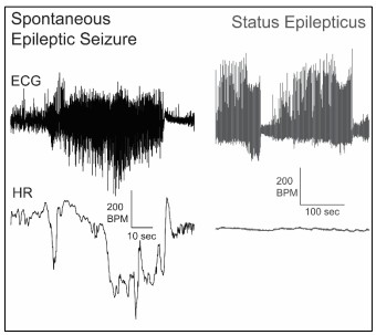
In Discussion: “Although the present study includes only non-fatal seizures, our report of HR during spontaneous seizure events supports work suggesting physiological events during non-fatal seizures predict SUDEP risk (Lamrani et al., 2023; Ryvlin, Nashef, & Tomson, 2013; Schuele et al., 2011). It remains to be determined if ictal events during fatal and non-fatal spontaneous seizures are different and future studies could help clarify any distinctions. Our examination focused on HR (and not respiration or BP). Whether exaggerated BJR-mediated HR response co-occurs with greater magnitude BJR-mediated apnea and hypotension remains to be determined.”
It is unclear if the bradycardias were spontaneous or a result of preceding central or obstructive apneas, oxygen desaturations, hypercapnia, arrhythmias, or other possible triggers.
Our work demonstrates the occurrence of ictal bradycardia whereas identifying precipitating factor(s) and interaction(s) of this phenomenon will require alternate approaches. Normal activation of hypoxic and hypercapnic ventilatory responses would be expected to increase HR. However, whether these chemoreflex circuits undergo remodeling in Kcc2/Crh mice remains unknown. Obstructive apnea via laryngospasm can cause reflex bradycardia, but we did not record airflow or respiratory EMG in these studies to determine the existence of obstructive apnea. More testing is merited.
In Discussion, “…Additional seizure-related disturbances such as central or obstructive apneas may contribute to BJR activation. Hypoxia, which could result from ictal apnea, is known to increase excitatory neurotransmission to cardiac vagal motor neurons that cause vagal bradycardia (Griffioen et al., 2007) and induces platelet activation (Tyagi et al., 2014) which is considered the main source of circulating serotonin for the BJR. As such, hypoxia resulting from apnea may increase likelihood of exaggerated BJR during seizures. Consistent with this…”
Considerable prior work in the literature suggests SUDEP could be mediated, in some patients, by a burst of parasympathetic activity to the heart. Were the heart rate changes in these animals during seizures inhibited or blocked by atropine or atenolol?
With this study targeting spontaneous seizures we were unable to test acute pre-treatment with atropine or atenolol. We did observe reduced mortality in Kcc2/Crh+KA mice that underwent chronic parasympathetic blockade via osmotic minipump of methylscopolamine (Figure 5).
In Discussion:
“Chronic inhibition of vagal parasympathetic motor output (the driver of BJR reflex bradycardia) improved mortality by 10% in Kcc2/Crh mice. Although this improvement provides some hope for patients at high risk for SUDEP with no treatment options, additional avenues of investigation are needed to more directly link seizure-related bradycardias to vagal parasympathetic motor output.”
The injection of the 5HT agonist phenylbiguanide into the right jugular is not a selective approach for activating the Bezold Jarisch Reflex (BJR), which is caused by increased activity of intracardiac sensory neurons (generally activated with is chemia or a combination of low preload with high contractility). The results should be interpreted more cautiously, as a response to systemic administration of phenylbiguanide only.
BJR can be experimentally triggered with intravenous infusion of various compounds including veratrum alkaloids (Cramer, 1915), 5HT (Fozard 1983), or 5HT3R agonists (Verberne & Guyenet, 1992). We now specifically refer to BJR in our study as that induced by PBG (a 5HT3R agonist), as others have done (Yamano et al., 1995; PMID: 8786638) and acknowledge endogenous BJR activation in the discussion.
Added to the results: “As dysfunction of serotonergic signaling is implicated in the pathophysiology of SUDEP (Richerson & Buchanan, 2011), we investigated the Bezold Jarisch Reflex (BJR) (Fig. 5), a cardioinhibitory reflex that is reliably triggered experimentally by activation of cardiopulmonary vagal afferents containing serotonin type 3 receptors (5HT3R) (Fozard 1983; Yamano et al., 1995).”
Added to Discussion: “Although our report is the first to link BJR to SUDEP, serum serotonin levels are elevated following generalized seizures (Murugesan et al., 2018), likely via release from activated platelets (Cloutier et al., 2018). This surge in serum serotonin could lead to endogenous activation of BJR, as bolus intravenous infusion of serotonin reliably triggers BJR experimentally (Fozard 1983; Whalen et al., 2000).”
Reviewer #2 (Public review):
Some of the conclusions may be a bit overstated as is and would benefit from more discussion and perhaps additional data.
Post-ictal HR data are now included in the Results, Table 2.1 and Figure 2.
The Discussion now includes more details regarding respiration, BP, obstructive apnea, properties of the BJR, and distinctions in seizure type based on whether evoked or lethal.



