
Peer review process
Not revised: This Reviewed Preprint includes the authors’ original preprint (without revision), an eLife assessment, public reviews, and a provisional response from the authors.
Read more about eLife’s peer review process.Editors
- Reviewing EditorAthena AkramiSainsbury Wellcome Centre, University College London, London, United Kingdom
- Senior EditorLaura ColginUniversity of Texas at Austin, Austin, United States of America
Reviewer #1 (Public review):
Summary:
In this study, the authors trained mice to perform a memory-guided navigation task, in which they must navigate to a previously cued arm after a delay period. They optogenetically inhibited the dorsal hippocampus or mPFC (targeting PL) in different task epochs. They found that for both regions, inactivating at the beginning of the navigation epoch impaired performance and induced mice to revert to habitual side biases. Inactivating during other epochs, including a delay period before the navigation phase, had little or no impact on behavior. The relationship between trial duration and behavioral performance was differentially impacted by hippocampal and mPFC inactivation, suggesting that the nature of the deficits was somewhat different.
Strengths:
The effects of perturbations are robust across animals and generally convincing. The lack of effect at some task epochs serves as a nice internal control. The finding that hippocampus and mPFC inactivation produced subtly different effects is interesting.
Weaknesses:
The simplicity of the behavior makes it difficult to resolve how exactly the hippocampus and mPFC contribute to working memory. Also, the language does not always reflect the trends in the data: the authors claim that optogenetic perturbations cause mice to repeat previous choices, but the data show that perturbations increase the likelihood of choosing a preferred side (which is left for most mice). A side bias is not the same as choice repetition. This has implications for interpreting the nature of the behavioral effects.
Reviewer #2 (Public review):
Summary:
The study uses transient optogenetic silencing of the dorsal hippocampus or prefrontal cortex in mice using a delayed response task in a T-maze, with a temporal delay of 1s after a visual cue, followed by a central stem run period before choice execution. Silencing of either the dorsal hippocampus or the dorsal prefrontal cortex is executed for varying time periods during the stem/ central arm running epoch or the 1s temporal delay epoch by targeting either PV+ or Dlx interneurons in a block-wise or random-trial design. The main result reported is that silencing of either region during long periods of stem running impaired choice behavior, and silencing during the temporal delay period did not have an effect.
Strengths:
The major strength of the study is using the optogenetic silencing strategy to target different temporal periods of the task.
Weaknesses:
(1) A major weakness of the study is the lack of a balanced design with equal time periods of silencing during the stem running period and temporal delay period in many of the animals, which precludes any conclusion about distinct functional roles of the regions during these two phases of the task. The central question of this study is not new, with many previous studies investigating distinct and overlapping roles of hippocampus and prefrontal cortex in spatial working memory tasks and memory-guided navigation, using inactivation of one or both regions, crossed inactivation approaches, as well as targeting direct and indirect connections between the regions (PMIDs: 20074655, 9030646, 17045348, 30179661, 27511010, 10491611, 26017312, etc.), in addition to several physiology studies. A key extension for the current study would have been to show a distinction between roles in the temporal delay period after the cue and the stem-running working memory period. However, the inactivation period during stem running shows effects only for long inactivation periods, 2s initial periods and later ~1.6s periods (run after 0.8s, >2/3 total running periods), whereas the temporal delay period inactivation is 1s for the large majority of animals, which is a clear mismatch in inactivation periods, obviating this conclusion of distinction.
(2) It is not clarified why such short delay periods were used compared to long periods of ~10s in T-maze spatial alternation tasks with delay, and whether the temporal delay period of 1 sec is strictly distinct from the spatiotemporal delay period during stem running in terms of short-term memory function. The choice of time windows for inactivation needs to be better justified, which currently appears to be rather random (2s initial running period, run after 0.8s, run after 1.6s; for an average reported running period of ~2.4-2.5s). The ideal design clearly would have been to use a 2s temporal delay period so that the inactivation time in this epoch matched the initial 2s run period. Only a subset of 4 animals were run with a longer temporal delay period, and too with only with hippocampal inactivation (Figure 4d). The main conclusion of distinction between temporal delay and stem running delay periods is therefore not adequately tested for the prefrontal cortex, and the statistics in terms of number of animals for this important control for hippocampal inactivation are also not comparable to the main experiment.
(3) The mixture of PV-Cre animals (5 animals), Dlx targeting (2 animals), and one WT animal is also suggestive of a fragmented approach, and inactivation efficacy cannot be assumed to be similar for different animals. Importantly, there is no physiological evidence for confirmation of suppression in the optogenetic experiments, even in exemplar animals.
Reviewer #3 (Public review):
The authors sought to determine when the dorsal hippocampus and mPFC are causally required during a delayed spatial working memory task. Using temporally precise optogenetic silencing in mice performing a delayed cue-guided T-maze task, they tested the effects of transient perturbations during distinct behavioral epochs. Contrary to the common view that these regions are primarily required during the delay period to maintain working memory representations, they report that silencing during the delay had little effect on performance, whereas perturbation during the early phase of central-arm traversal consistently impaired performance. The authors conclude that hippocampal and prefrontal contributions to memory-guided behavior are dynamically engaged during active navigation rather than passive maintenance of information.
The study has several strengths. The behavioral paradigm is carefully designed to dissociate cue, delay, and movement epochs, allowing temporally specific causal manipulations. The systematic comparison of multiple task epochs represents a major strength and provides compelling evidence that the behavioral effects of perturbation are epoch-dependent. The authors also include several important controls, including stimulation during multiple task phases, a longer-delay condition to dissociate task epoch from elapsed time since cue presentation, and analyses of movement trajectories and perseverative behavior that provide additional insight into the nature of the behavioral deficits. Together, these experiments convincingly demonstrate that transient dorsal hippocampal and mPFC perturbations have markedly different behavioral consequences depending on when they occur within a trial.
The evidence is generally solid and supports the primary finding that perturbations during early navigation produce larger impairments than perturbations during the delay period. However, some aspects of the broader interpretation are less well supported. Most notably, the study lacks a non-memory control task, such as a visually guided version of the maze, making it difficult to determine whether the observed deficits specifically reflect disruption of memory-guided behavior or more general impairments in action selection, behavioral flexibility, or movement planning. The observed increase in perseverative responding and delayed commitment to a turn are consistent with either interpretation. In addition, several experimental conditions rely on relatively small numbers of animals, limiting confidence in some negative findings, particularly for the longer-delay and later-run manipulations. Finally, while the Discussion proposes that hippocampal-prefrontal circuits become engaged during the transformation of stored information into action, this mechanistic interpretation remains speculative because no neural recordings accompany the causal manipulations.
Overall, the authors achieve their primary aim of demonstrating that the behavioral consequences of dorsal hippocampal and mPFC silencing depend strongly on task epoch. The data convincingly support the conclusion that these structures are more vulnerable to perturbation during early navigation than during the brief delay period used in this task. The broader conclusion that these findings redefine when hippocampal-prefrontal circuits support working memory should be interpreted more cautiously, as alternative explanations involving action selection or behavioral state remain plausible in the absence of additional control tasks.
The findings are potentially important because they challenge the common assumption that hippocampal and prefrontal contributions to delayed-response tasks are centered on delay-period maintenance. Instead, the work supports the idea that these circuits may be recruited when remembered information is translated into goal-directed behavior. This framework is broadly consistent with recent distributed models of working memory and provides an interesting perspective that may help reconcile previous studies reporting effects during different task phases. The behavioral paradigm and temporally precise perturbation approach should also be useful for future studies aimed at dissecting the dynamic contributions of hippocampal-prefrontal circuits during memory-guided behavior.
Author response:
On the eLife Assessment. We appreciate the assessment’s recognition that this study addresses a long-standing question concerning hippocampal and prefrontal contributions to working memory. We think the broader significance lies in constraining how causal manipulations in working-memory tasks are interpreted. Working memory encompasses many processes distributed across a trial and showing that a region is required for a delayed-response task does not establish when its contribution is necessary. The observation that hippocampus and mPFC are required while navigating to a goal, but not during stationary delay, challenges a common assumption and, in our opinion, has implications across the broader field of working-memory research.
We agree that the original manuscript did not adequately report effect sizes or convey the uncertainty associated with some smaller samples. However, the dataset does include duration-matched stationary and running conditions, including a hippocampal long-delay control matched to early-running stimulation in both duration and elapsed time after cue onset. The new effect-size and within-animal analyses support a robust hippocampal epoch difference, while the corresponding mPFC comparison is less precisely estimated and should be interpreted more cautiously.
We therefore think the central finding remains well supported: hippocampal function, and potentially mPFC function, is required during the active navigation period of this memory-guided task but not detectably during the stationary delay. This does not establish the specific computation disrupted during running. Neural recordings would certainly provide further insight into the underlying mechanism, but their absence does not detract from the value of the behavioral result itself.
Reviewer #1 (Public review):
The simplicity of the behavior makes it difficult to resolve how exactly the hippocampus and mPFC contribute to working memory.
The task was designed to combine the temporal precision of cue-based delayed-response paradigms with the behavioral richness of freely moving navigation. Few tasks combine a fixed cue-presentation period, an explicit delay, and a subsequent navigation phase involving extended running. This structure creates well-defined behavioral epochs that can be targeted with temporally precise perturbations, allowing us to ask when hippocampal and mPFC contributions are required within an ongoing memory-guided behavior. How these regions contribute is the harder question, and one we are pursuing next. Identifying when perturbations disrupt behavior is an important step toward understanding how these regions support memory-guided navigation
Also, the language does not always reflect the trends in the data: the authors claim that optogenetic perturbations cause mice to repeat previous choices, but the data show that perturbations increase the likelihood of choosing a preferred side (which is left for most mice). A side bias is not the same as choice repetition. This has implications for interpreting the nature of the behavioral effects.
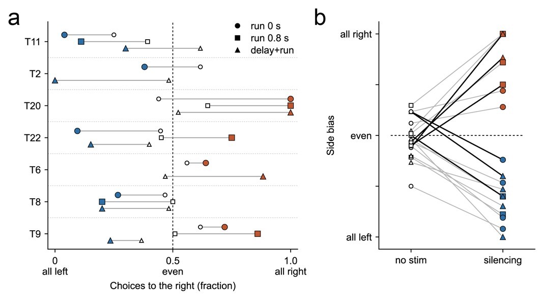
We agree that our results do not clearly distinguish a directional bias from a tendency to repeat the previous choice. To examine whether mice consistently favored a particular direction, we compared their side preferences during silencing across sessions. Mice generally favored the same side across silencing conditions, although some switched direction in individual sessions (Author response image 1a). Within sessions, the preferred side was maintained from no-stimulation to stimulation trials in 13 of 19 cases and reversed in six (Author response image 1b). These observations are consistent with a directional preference that can sometimes reverse during stimulation, and cannot be disambiguated from perseveration. In the revision, we will describe the effect as increased directional bias and revise the language concerning choice repetition and perseveration throughout the manuscript.
Author response image 1.
Silencing increases directional bias. (a) Fraction of choices made to the right in each session, for every mouse (rows) and each silencing condition (symbols). Open symbols, no-stimulation trials; filled symbols, stimulation trials from the same session; blue and red denote a left or right preference during silencing. Filled symbols falling predominantly on the same side of 0.5 within a row indicate that a mouse generally favored the same direction across silencing conditions, although some mice switched direction. One session per mouse and condition, hippocampal silencing only; T2 and T6 did not perform the 0.8 s condition. (b) The same sessions expressed as signed bias, from no stimulation to silencing. Black lines mark the six sessions in which the preferred side reversed; grey lines the thirteen in which it was maintained.
Reviewer #2 (Public review):
A major weakness of the study is the lack of a balanced design with equal time periods of silencing during the stem running period and temporal delay period in many of the animals, which precludes any conclusion about distinct functional roles of the regions during these two phases of the task. The main conclusion of distinction between temporal delay and stem running delay periods is therefore not adequately tested for the prefrontal cortex, and the statistics in terms of number of animals for this important control for hippocampal inactivation are also not comparable to the main experiment.
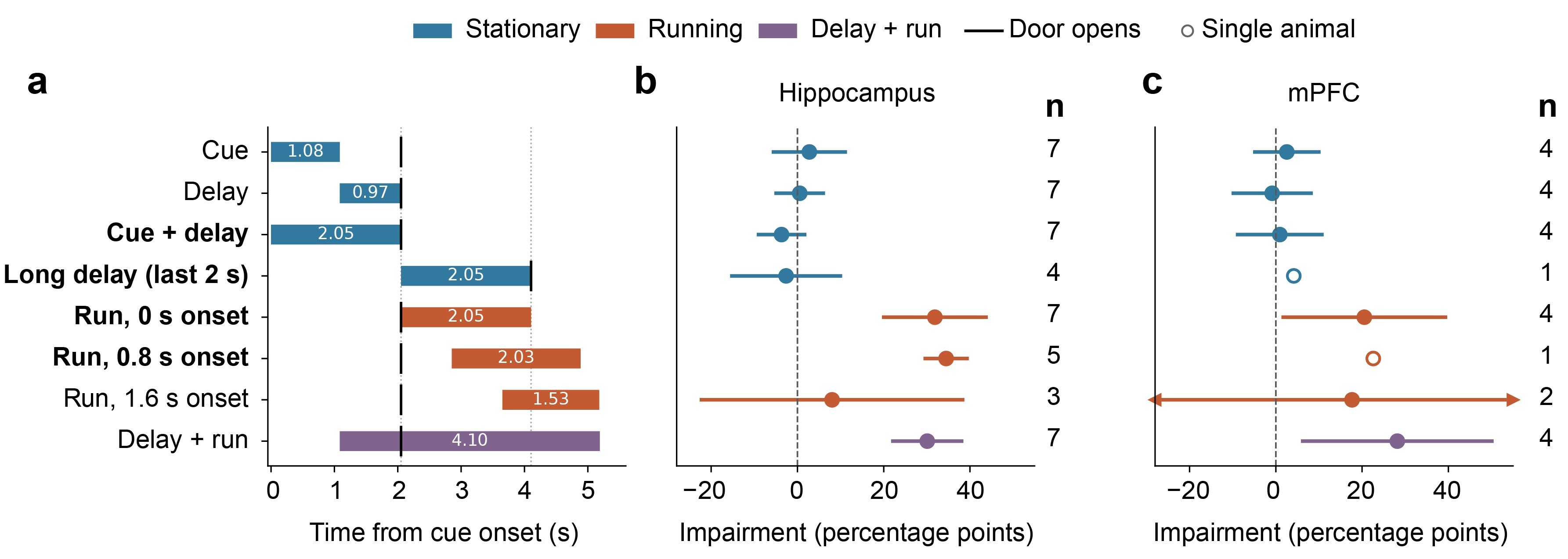
We agree that matching stimulation duration is an essential control and recognize that the relevant comparisons and statistics were not sufficiently clear in the original manuscript. Four hippocampal conditions used closely matched stimulation durations of 2 s: Cue+Delay, long delay, early running, and running with a 0.8 s onset (Author response image 2a). Crucially, the long-delay control matched both stimulation duration and elapsed time after cue onset to the early-run condition, while the mouse remained stationary.
Author response image 2b–c shows the estimated impairment and its 95% confidence interval for each condition. In the hippocampal experiments, the duration-matched stationary conditions showed effects close to zero, whereas the running conditions showed large impairments. Despite the smaller sample, the upper confidence limit for the long-delay impairment was approximately 10 percentage points, substantially below the observed early-running impairment. These estimates establish that despite the smaller sample, the data support a lack of effect compared to early running.
Author response image 2.
Duration-matched stimulation produces different behavioral effects across task epochs. (a) Stimulation timing relative to cue onset. Numbers within bars indicate calculated median stimulation duration in seconds; black ticks indicate door opening. Bold labels identify conditions with 2 s stimulation. (b-c) Mean impairment in choice accuracy for hippocampal and mPFC manipulations. Impairment is accuracy during baseline minus accuracy with stimulation, in percentage points. Error bars show 95% confidence intervals across animals; numbers indicate mice. Open circles denote single-animal observations, and arrows indicate confidence intervals extending beyond the plotted range.
For mPFC, the duration-matched Cue+Delay condition likewise showed an effect close to zero, whereas early-running stimulation produced substantial impairment. However, the long-delay condition included only one mouse. The later-running effects in both regions were also less precisely estimated. We will distinguish these limitations from the more informative stationary-condition results.
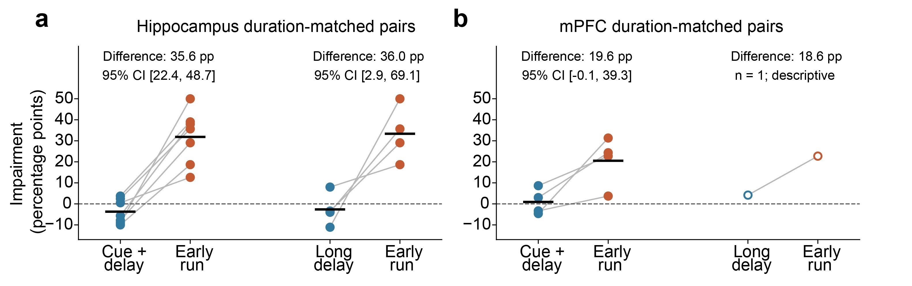
To directly test whether the duration-matched effects differed across epochs, we next compared impairment within the same mice, including only animals tested in both conditions (Author response image 3). For the hippocampus, every mouse showed greater impairment during early running than during either duration-matched stationary condition, and the confidence intervals for both paired differences excluded zero. These within-animal comparisons support an epoch-dependent effect that cannot be explained by stimulation duration alone. The mPFC comparison showed the same direction of effect, although the confidence interval for the Cue+Delay versus running difference narrowly included zero. We will therefore distinguish the stronger evidence for the hippocampal epoch difference from the more limited evidence for mPFC.
Author response image 3.
Within-animal comparisons of duration-matched stimulation effects. (a–b) Impairment during Cue+Delay or long-delay stimulation compared with early-running stimulation for hippocampal (a) and mPFC (b) manipulations. Points represent individual mice, lines connect observations from the same mouse, and black bars indicate mean. Annotations report the mean paired difference in impairment (running minus stationary), and its 95% confidence interval calculated using the t distribution. Positive differences indicate greater impairment during running. The single-mouse mPFC long-delay comparison is descriptive. Blue indicates stationary epochs and orange indicates running.
The central question of this study is not new, with many previous studies investigating distinct and overlapping roles of hippocampus and prefrontal cortex in spatial working memory tasks and memory-guided navigation, using inactivation of one or both regions, crossed inactivation approaches, as well as targeting direct and indirect connections between the regions (PMIDs: 20074655, 9030646, 17045348, 30179661, 27511010, 10491611, 26017312, etc.), in addition to several physiology studies.
We agree that hippocampal and prefrontal contributions to spatial working memory have been extensively studied. That’s precisely why we find these results impactful when placed in the rich context of the field. The requirement for these regions in delayed working memory tasks has often been interpreted in terms of their contributions during the delay period. However, a requirement during a delay-based task does not itself demonstrate a requirement during the delay. Previous manipulations have not isolated delay periods of waiting from the subsequent navigation within a trial.
Our experiments extend this work by separately targeting cue presentation, the delay, and different portions of navigation within the same task. This allows us to test whether the behavioral consequences of perturbation depend on the particular epoch in which it occurs. This distinction is important: knowing that a region is required for a memory-guided task does not establish when its contribution is needed. Identifying those periods constrains how we interpret the deficits produced by longer-lasting inactivation. We believe that this is an important result that should be considered when interpreting these broader findings. We will ensure that the appropriate literature and discussion are included in the revision.
It is not clarified why such short delay periods were used compared to long periods of ~10s in T-maze spatial alternation tasks with delay, and whether the temporal delay period of 1 sec is strictly distinct from the spatiotemporal delay period during stem running in terms of short-term memory function.
The 1 s delay was chosen to maintain reliable task performance, as some mice could not perform the task with longer delays. Indeed, one reason the longer-delay condition includes fewer animals is that two mice could not perform reliably with the 3 s delay (one additional mouse was not tested in this condition). Delays on this timescale have also been used in rodent cued delayed-response tasks, including a 0.5 s delay in Kopec et al. (2015), a 1.3 s delay in Guo et al. (2014), and a 1.2 s delay in Inagaki et al. (2019). Like these tasks, our paradigm requires mice to remember an externally presented cue specifying the upcoming response, rather than their own previous arm choice as in spatial alternation. It therefore combines spatial navigation with a cued delayed-response requirement, and the delay durations tolerated in alternation tasks are not necessarily directly comparable. We will clarify this rationale in the manuscript.
We agree that the stationary delay and the subsequent run both require retention of information after cue offset. Our experiments distinguish these behavioral epochs, but do not establish that they involve separate short-term memory processes. The different effects of perturbation suggest that the contribution of these regions changes as the animal moves from waiting to navigating. Determining what accounts for this change is an important direction for future work.
The choice of time windows for inactivation needs to be better justified, which currently appears to be rather random (2s initial running period, run after 0.8s, run after 1.6s; for an average reported running period of ~2.4-2.5s).
We sought to target different portions of the central-arm run. Given the typical traversal time of approximately 2.4–2.5 s, stimulation onsets at 0, 0.8, and 1.6 s sampled the beginning, middle, and later portions of the run. Our setup allowed precise control of stimulation timing, and, as shown in Figure 4b, these onset times correspond approximately to the start, middle, and end of the central arm. Each condition used a nominal 2 s stimulation window, truncated if the mouse reached the choice point sooner. The windows therefore overlap, with the later-onset condition generally producing shorter stimulation. We will clarify this rationale and the distinction between stimulation onset and duration in the revised manuscript.
The mixture of PV-Cre animals (5 animals), Dlx targeting (2 animals), and one WT animal is also suggestive of a fragmented approach, and inactivation efficacy cannot be assumed to be similar for different animals. Importantly, there is no physiological evidence for confirmation of suppression in the optogenetic experiments, even in exemplar animals.
The Dlx animals were included to improve regional specificity through local viral expression and to test whether the behavioral effects were consistent across targeting approaches. Both approaches produced comparable impairments during running, supporting their inclusion in the same analysis. We will clarify this rationale in the manuscript.
Optogenetic activation of inhibitory interneurons is an established approach for suppressing local principal-cell activity, with physiological validation in previous studies (Guo et al., 2014; Li et al., 2019, Zutshi et al., 2022). In our experiments, running-period stimulation produced robust behavioral impairments that were consistent across animals and targeting approaches. Furthermore, comparisons across epochs were performed within animals, using the same preparation and stimulation parameters. Differences in efficacy between animals therefore cannot readily account for the observed epoch dependence. Although direct recordings would establish the magnitude and spatial extent of suppression in our preparation, the central behavioral finding is supported by these within-animal comparisons.
Reviewer #3 (Public review):
The findings are potentially important because they challenge the common assumption that hippocampal and prefrontal contributions to delayed-response tasks are centered on delay-period maintenance.
We thank the reviewer for describing our findings as “potentially important” and for highlighting their implications for how hippocampal and prefrontal contributions to delayed-response tasks are understood. We appreciate the constructive suggestions and address the public comments below.
Most notably, the study lacks a non-memory control task, such as a visually guided version of the maze, making it difficult to determine whether the observed deficits specifically reflect disruption of memory-guided behavior or more general impairments in action selection, behavioral flexibility, or movement planning. The observed increase in perseverative responding and delayed commitment to a turn are consistent with either interpretation.
We agree that leaving the cue on throughout the trial would provide an important control for distinguishing memory-specific effects from broader effects on action selection or movement planning. The senior author is currently setting up a new laboratory, so implementing this control may take some time. We hope to include it in the revised manuscript.
The running-period deficit indicates a disruption of processes that enable the animal to act on a remembered cue. Whether this reflects disruption of memory itself, movement planning, or another component of translating the cue into a choice requires further clarification but does not detract from the observed dependence on task epoch. Uncertainty about the mechanism of the running-period deficit also does not change the observation that the same manipulation produced no detectable impairment during the stationary delay, when the cue was absent and still had to be remembered. This will be clearly discussed in the revision.
In addition, several experimental conditions rely on relatively small numbers of animals, limiting confidence in some negative findings, particularly for the longer-delay and later-run manipulations.
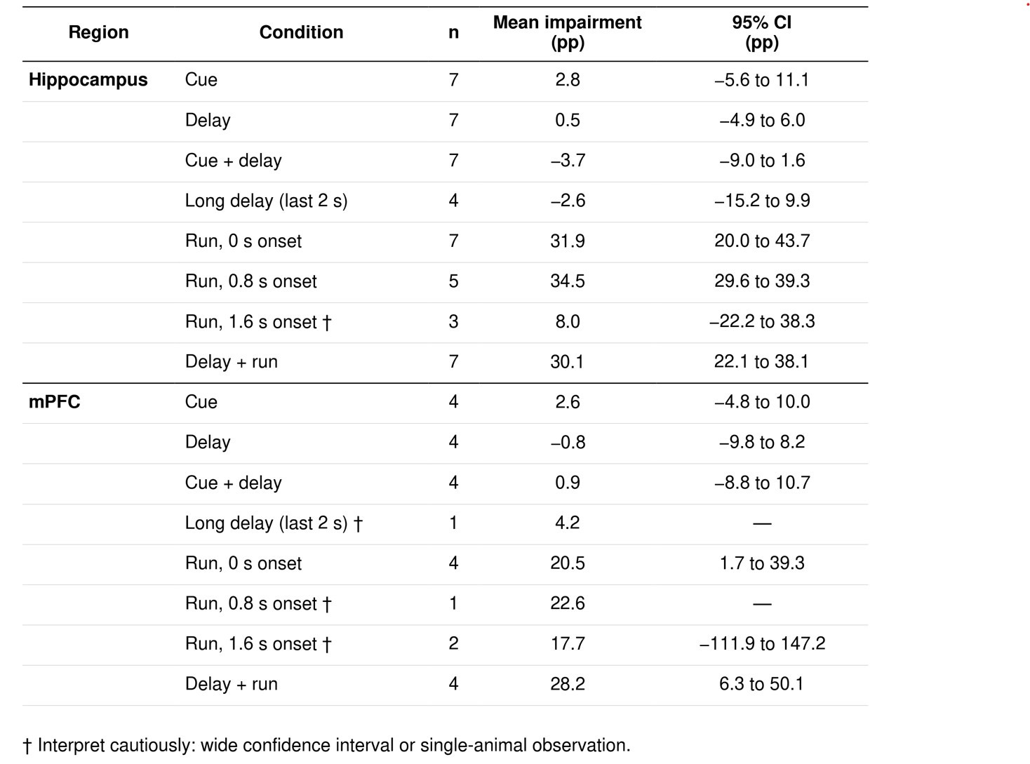
We agree that small sample sizes limit the interpretation of some negative findings. We now report animal-level effect estimates and 95% confidence intervals for each condition (Author response image 2b–c; Author response table 1).
Of the nine conditions with no detectable impairment and more than one mouse, seven had confidence intervals that excluded effects as large as the observed mean early-running impairment in the same region. This included the hippocampal long-delay condition, despite its smaller sample. These results argue against similarly large impairments in these conditions, although smaller effects remain possible. The two later-run conditions remained too uncertain to exclude such impairments. These and the two single-animal conditions are marked in Author response table 1 and will be interpreted cautiously.
Author response table 1.
Animal-level impairment estimates and uncertainty
Finally, while the Discussion proposes that hippocampal-prefrontal circuits become engaged during the transformation of stored information into action, this mechanistic interpretation remains speculative because no neural recordings accompany the causal manipulations.
We will clarify in the Discussion that the proposed transformation of stored information into action remains speculative. Nevertheless, we believe this is an exciting possibility raised by our findings that warrants further investigation. Neural recordings would help test this interpretation but are beyond the scope of the current paper. We plan to explore this question in future work.
References
Guo ZV, Li N, Huber D, Ophir E, Gutnisky D, Ting JT, Feng G, Svoboda K (2014). Flow of cortical activity underlying a tactile decision in mice. Neuron 81:179–94. doi:10.1016/j.neuron.2013.10.020. PMID 24361077.
Kopec CD, Erlich JC, Brunton BW, Deisseroth K, Brody CD (2015). Cortical and subcortical contributions to short-term memory for orienting movements. Neuron 88:367–77. doi:10.1016/j.neuron.2015.08.033. PMID 26439529.
Inagaki HK, Fontolan L, Romani S, Svoboda K (2019). Discrete attractor dynamics underlies persistent activity in the frontal cortex. Nature 566:212–217. doi:10.1038/s41586-019-0919-7. PMID 30728503.
Li N, Chen S, Guo ZV, Chen H, Huo Y, Inagaki HK, Chen G, Davis C, Hansel D, Guo C, Svoboda K (2019). Spatiotemporal constraints on optogenetic inactivation in cortical circuits. eLife 8:e48622. doi:10.7554/eLife.48622. PMID 31736463.
Zutshi I, Valero M, Fernández-Ruiz A, Buzsáki G (2022). Extrinsic control and intrinsic computation in the hippocampal CA1 circuit. Neuron 110:658–673.e5. doi:10.1016/j.neuron.2021.11.015. PMID 34890566.



