
Peer review process
Revised: This Reviewed Preprint has been revised by the authors in response to the previous round of peer review; the eLife assessment and the public reviews have been updated where necessary by the editors and peer reviewers.
Read more about eLife’s peer review process.Editors
- Reviewing EditorKevin EadeLowy Medical Research Institute, La Jolla, United States of America
- Senior EditorLori SusselUniversity of Colorado Anschutz Medical Campus, Aurora, United States of America
Reviewer #1 (Public review):
Bajohr and colleagues propose a transcription factor-driven approach to generating bonafide oligodendrocyte lineage cells (OLCs) from primary mouse astrocytes. Ectopic expression of Olig2, Sox10, or Nkx6.2 in isolated astrocytes produced a range of OLC-like cell states, with Sox10 emerging from lineage tracing and single cell RNA sequencing experiments as the most successful transcription factor in driving direct lineage reprogramming. The authors strengthened their claims with an unbiased, deep learning perturbation model to predict genetic drivers of the astrocyte cluster to OLC cluster transition observed in their scRNA seq dataset. Here, Sox10 surfaced in the top ten correlated genes, and the top transcription factor, mediating this fate shift. Altogether, this paper presents an interesting approach to generate OLCs, a cell type historically difficult to procure, from primary mouse astrocytes to study this lineage in development and disease and perhaps repopulate it in dysmyelinating conditions. While this certainly addresses a technical gap in the field, authors defined iOLCs as ones with lineage-specific gene expression and morphological characteristics, lacking any functional analysis to assess the reprogrammed cells' capacity to myelinate. This comment and other critiques are discussed below.
While Sox10 and Mbp expression in iOLCs, as confirmed by IHC, is a promising result suggesting that ectopic Sox10 instructs transduced cells to develop into cells of myelinating potential, functional confirmation is essential. As mentioned in the discussion, the absence of a substrate for myelination may have also contributed to the low DLR efficiency. Co-culturing Sox10 iOLCs with primary neurons and examining the cells' potential to engage and enwrap axons would greatly strengthen the authors' claim that this could be an effective therapeutic approach to myelin regeneration in vivo, or even a technical approach to studying myelin dynamics in vitro.
In Figure 1B, it appears that Mbp expression in tdTomato+ cells decreases in Sox10 transduced iOLs during the observed time period. Can the authors elaborate on this result, given that MBP expression is crucial for myelination and should, if anything, increase with time?
The authors acknowledge that there is a conversion of tdTomato- zsGreen+ cells with an astrocyte-like morphology to OLC cells expressing Mbp following Sox10 induction (Supplementary figure 5C,D). While they note the diversity of the astrocyte lineage in the discussion, further analysis should be applied to this subset of cells to confirm the subset of astrocyte or progenitor-like cell type that gives rise to their cell endpoint of interest (Sox10-driven Mbp+ iOLs).
Finally, ectopic expression of Olig2 and Sox10 in primary astrocytes resulted in very different OLC subtypes, as evidenced by OLC marker expression seen in IHC and the subclustering of these cell types in scRNA seq. Although this diversity in OLC type and generation efficiency follows with previous reports showing that these two transcription factors vary in effect, might the authors further discuss this discrepancy given that the two transcription factors regulate one another (as mentioned in the introduction) and should theoretically give rise to more similar cells? Perhaps due to the lower specificity of Olig2 in marking a pure OLC population relative to Sox10?
Author response:
The following is the authors’ response to the previous reviews.
Public Reviews:
Reviewer #1 (Public review):
Bajohr and colleagues propose a transcription factor-driven approach to generating bonafide oligodendrocyte lineage cells (OLCs) from primary mouse astrocytes. Ectopic expression of Olig2, Sox10, or Nkx6.2 in isolated astrocytes produced a range of OLC-like cell states, with Sox10 emerging from lineage tracing and single cell RNA sequencing experiments as the most successful transcription factor in driving direct lineage reprogramming. The authors strengthened their claims with an unbiased, deep learning perturbation model to predict genetic drivers of the astrocyte cluster to OLC cluster transition observed in their scRNA seq dataset. Here, Sox10 surfaced in the top ten correlated genes, and the top transcription factor, mediating this fate shift. Altogether, this paper presents an interesting approach to generate OLCs, a cell type historically difficult to procure, from primary mouse astrocytes to study this lineage in development and disease and perhaps repopulate it in dysmyelinating conditions. While this certainly addresses a technical gap in the field, authors defined iOLCs as ones with lineage-specific gene expression and morphological characteristics, lacking any functional analysis to assess the reprogrammed cells' capacity to myelinate. This comment and other critiques are discussed below.
While Sox10 and Mbp expression in iOLCs, as confirmed by IHC, is a promising result suggesting that ectopic Sox10 instructs transduced cells to develop into cells of myelinating potential, functional confirmation is essential. As mentioned in the discussion, the absence of a substrate for myelination may have also contributed to the low DLR efficiency. Co-culturing Sox10 iOLCs with primary neurons and examining the cells' potential to engage and enwrap axons would greatly strengthen the authors' claim that this could be an effective therapeutic approach to myelin regeneration in vivo, or even a technical approach to studying myelin dynamics in vitro.
In Figure 1B, it appears that Mbp expression in tdTomato+ cells decreases in Sox10 transduced iOLs during the observed time period. Can the authors elaborate on this result, given that MBP expression is crucial for myelination and should, if anything, increase with time?
The authors acknowledge that there is a conversion of tdTomato- zsGreen+ cells with an astrocyte-like morphology to OLC cells expressing Mbp following Sox10 induction (Supplementary figure 5C,D). While they note the diversity of the astrocyte lineage in the discussion, further analysis should be applied to this subset of cells to confirm the subset of astrocyte or progenitor-like cell type that gives rise to their cell endpoint of interest (Sox10-driven Mbp+ iOLs).
Finally, ectopic expression of Olig2 and Sox10 in primary astrocytes resulted in very different OLC subtypes, as evidenced by OLC marker expression seen in IHC and the subclustering of these cell types in scRNA seq. Although this diversity in OLC type and generation efficiency follows with previous reports showing that these two transcription factors vary in effect, might the authors further discuss this discrepancy given that the two transcription factors regulate one another (as mentioned in the introduction) and should theoretically give rise to more similar cells? Perhaps due to the lower specificity of Olig2 in marking a pure OLC population relative to Sox10?
We thank the editor and reviewers for their additional comments, which have significantly improved our manuscript.
Recommendations for the authors:
Reviewer #1 (Recommendations for the authors):
The authors use Aldh1l1+ astrocyte with a GFAP promoter linked to TF expression, claiming that the converting cells are cortical astrocytes. However, during early mouse development, radial glia that can give rise to multiple cell types, including oligodendrocytes, also express Aldh1l1 and low levels of GFAP. Therefore, it is not proven whether the resulting iOLs came from mature astrocyte or a radial glia population. Especially since in Figure 3B,G it is shown that a majority of the D0 population expresses high amounts of Vimentin and Nestin, both markers associated with radial glia and immature astrocytes. It would be beneficial for the authors to confirm that either there are no contaminating radial glia or that the radial glia don't express the TF, especially since the iOL population is so small.
Thank you for this comment. We agree that previous studies have demonstrated Aldh1l1 expression in radial glia cells [1]. However, our dissection protocol to obtain the postnatal astrocytes is cortex-specific and does not take portions of the VZ/SVZ, preventing radial glia contamination.
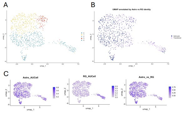
Nevertheless, to confirm the astrocytic identity of our Aldh1l1+ starting cells we used AUCell enrichment scoring [2]. First, Aldh1l1+ cells were subclustered from our starting culture single cell dataset (Author response image 1A). We then defined two gene signature modules: an “astrocyte” module, comprised of canonical astrocyte markers (Aqp4, Gja1, Slc1a2, Glul, Aldoc, S100b, Nfia, Thbs1, Cst3, Clu), and a “radial glia” module, containing common radial glia and progenitor markers (Pax6, Fabp7, Sox2, Hes1, Prom1, Top2a, Mki67, Ube2c, Cdk6, Mcm2). When we scored all Aldh1l1+ cells (n=869) for enrichment of each signature, no cells were classified as radial glia (Author response image 1B). Instead, the astrocyte signature predominated, with 68.3% classified as astrocytes (Author response image 1B,C). The remaining cells (36.7%), were classified as transitional, reflecting substantial expression of genes from both modules (Author response image 1B,C). Therefore, although the Aldh1l1 astrocytes do express genes common to radial glia, there are no cells that express only progenitor markers. This is consistent with literature showing that many radial glia genes are commonly found in astrocytes [3], [4], [5].
Taken together, our stringent dissection protocol and bioinformatic profiling of our starting Aldh1l1 cells suggests that the resulting Aldh1l1+iOLs are originating from astrocytes, rather than radial glia.
In Figure 2D, Sox10 and Nkx6.2 have an n=4 while the Cre control has an n=3. Why is this the case? Was the 4th point excluded? Similar attention should be given to other panels in Figure 2 for consistency.
We thank the reviewer for highlighting this. No outliers or datapoints were excluded from the analysis. Rather, the fourth culture for our control treated cells was not viable for analysis.
Author response image 1.
Astrocyte gene expression in Aldh1l1+ cells. (A) UMAP clustering of Aldh1l1+ cells in our starting cultures (0DPT, non-transduced). (B) UMAP clustering from (A) overlayed with cell classification based on AUCell gene signature expression scoring. (C) Feature plots showing AUCell enrichment scores (darker purple indicates higher enrichment) for the astrocyte signature (left), radial glia signature (middle), and the differential enrichment score (right, Astro_AUCell - RG_AUCell) (darker purple scores indicate higher astrocyte signature and negative scores (gray) indicate higher radial glia signature).
Representative images in Figure 2 do not convincingly support the argument by the authors. It appears that some of the cells highlighted by the arrows are just background (e.g. PDGFRa and td Tomato in Figure 2E, or zsGreen in Figure 2F). Additionally, the authors should show a different representative image depicting astrocyte morphology in Figure 2G 7DPT.
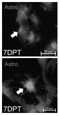
Thank you to the reviewer for this comment. We have replaced the images in Figure 2E,F to better represent our findings (updated manuscript Figure 2E,F). We have also adjusted the representative image in Figure 2G 7DPT to better visualize the astrocyte morphology (updated manuscript Figure 2G) as well as included as supplementary additional examples of pre-conversion astrocyte morphology to supplement our morphology analysis (Author response image 2).
Author response image 2.
Lineage tracing confirms true conversion of astrocytes to oligodendrocyte lineage cells. Representative images of astrocyte morphology observed prior to cell conversion (arrow indicates converting cells, scale bar =50um).
References
(1) L. C. Foo and J. D. Dougherty, “Aldh1L1 is expressed by postnatal neural stem cells in vivo,” Glia, vol. 61, no. 9, pp. 1533–1541, Sep. 2013, doi: 10.1002/glia.22539.
(2) S. Aibar et al., “SCENIC: Single-cell regulatory network inference and clustering,” Nat Methods, vol. 14, no. 11, pp. 1083–1086, Nov. 2017, doi: 10.1038/nmeth.4463.
(3) M. Götz and Y.-A. Barde, “Radial Glial Cells: Defined and MajorIntermediates between EmbryonicStem Cells and CNS Neurons,” Neuron, vol. 46, no. 3, pp. 369– 372, May 2005, doi: 10.1016/j.neuron.2005.04.012.
(4) P. Malatesta, I. Appolloni, and F. Calzolari, “Radial glia and neural stem cells,” Cell and Tissue Research, vol. 331, no. 1, pp. 165–178, 2008, doi: 10.1007/s00441-0070481-8.
(5) S. Clavreul, L. Dumas, and K. Loulier, “Astrocyte development in the cerebral cortex: Complexity of their origin, genesis, and maturation,” Front Neurosci, vol. 16, p. 916055, Sep. 2022, doi: 10.3389/fnins.2022.916055.



